. 2010 Nov;34(11):1879-87.
doi: 10.1111/j.1530-0277.2010.01276.x.

Fetal alcohol exposure increases mammary tumor susceptibility and alters tumor phenotype in rats

Affiliations

Fetal alcohol exposure increases mammary tumor susceptibility and alters tumor phenotype in rats

Tiffany A Polanco et al. Alcohol Clin Exp Res. 2010 Nov.

Abstract

Background: Altered fetal programming because of a suboptimal in utero environment has been shown to increase susceptibility to many diseases later in life. This study examined the effect of alcohol exposure in utero on N-nitroso-N-methylurea (NMU)-induced mammary cancer risk during adulthood.

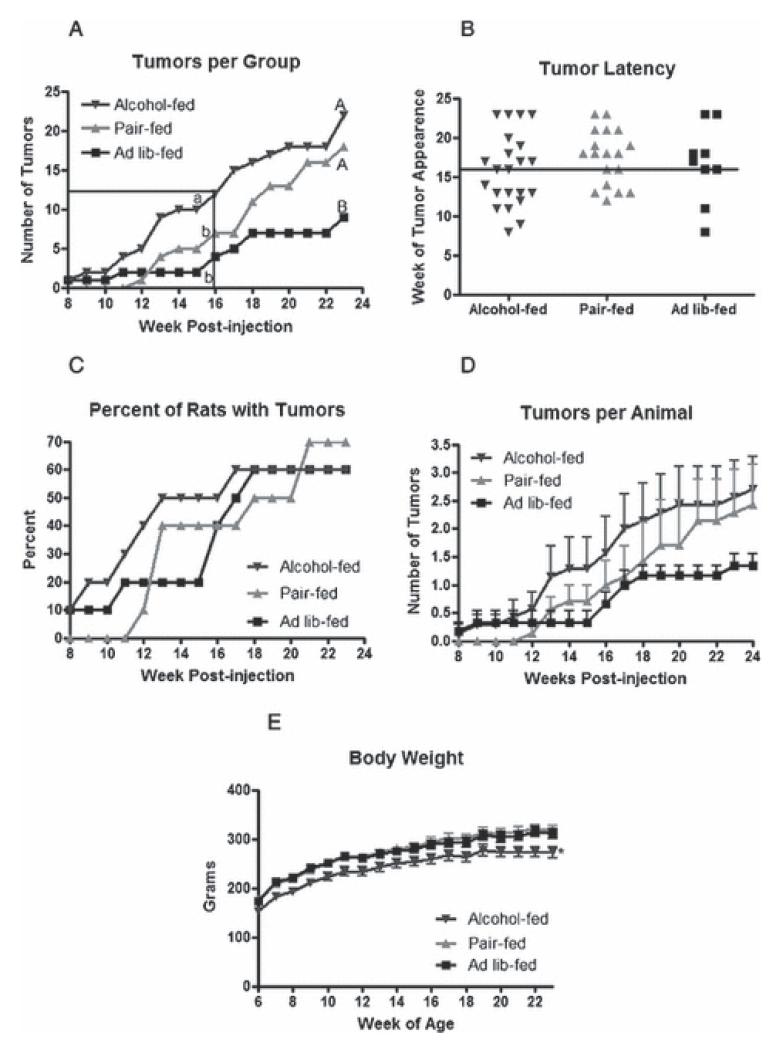
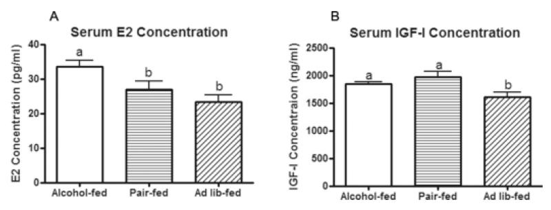
Methods: Study 1: Pregnant Sprague Dawley rats were fed a liquid diet containing 6.7% ethanol (alcohol-fed), an isocaloric liquid diet (pair-fed), or rat chow ad libitum (ad lib-fed) from day 11 to 21 of gestation. At birth, female pups were cross-fostered to ad lib-fed control dams. Adult offspring were given an I.P. injection of NMU at a dose of 50 mg/kg body weight. Mammary glands were palpated for tumors twice a week, and rats were euthanized at 23 weeks postinjection. Study 2: To investigate the role of estradiol (E2), animals were exposed to the same in utero treatments but were not given NMU. Serum was collected during the preovulatory phase of the estrous cycle.

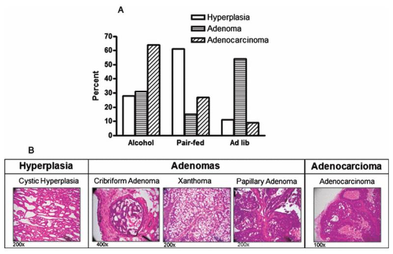
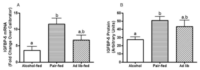
Results: At 16 weeks postinjection, overall tumor multiplicity was greater in the offspring from the alcohol-fed group compared to the control groups, indicating a decrease in tumor latency. At study termination, 70% of all animals possessed tumors. Alcohol-exposed animals developed more malignant tumors and more estrogen receptor-α-negative tumors relative to the control groups. In addition, IGF-binding protein-5 (IGFBP-5) mRNA and protein were decreased in tumors of alcohol-exposed animals. Study 2 showed that alcohol-fed animals had significantly increased circulating E2 when compared to either control group.

Conclusions: These data indicate that alcohol exposure in utero increases susceptibility to mammary tumorigenesis in adulthood and suggest that alterations in the IGF and E2 systems may play a role in the underlying mechanism.


Figures

Figure 1
Figure 1
Alcohol exposure in utero enhances mammary tumorigenesis. Animals exposed to alcohol in utero and control rats not exposed were administered a single I.P. injection of NMU at 50 mg/kg at approximately 50 days of age. Rats were palpated for tumors twice a week following injections. (a) Total number of tumors per treatment group each week post NMU injection. The boxed area represents when 50% tumor incidence was reached across all groups. Two time intervals were analyzed: 8-16 weeks (lower case letters, P < 0.05, n=10), and 8-23 weeks (upper case letters, P < 0.05, n=10). (b) Week of tumor appearance. Each data point represents an individual tumor. The horizontal line at week 16 post-injection represents when 50% tumor incidence occurred. (c) Percent of rats presenting with tumors each week post NMU injection, P = 0.9494, n=10. (d) Average number of tumors per animal in each group ± SEM. (e) Average body weight ± SEM with * denoting a significant difference (P < 0.005, n=10). Data in panels a, d, and e were analyzed using a Kruskal-Wallis non-parametric one way ANOVA with a Dunn’s Multiple Comparison post-hoc analysis at the level of α = 0.05, and panel c was analyzed using a log-rank test.
Figure 2
Figure 2
Alcohol exposure in utero results in altered tumor development. Tumors were hematoxylin and eosin stained for histological evaluation. (a) Percent of tumors of each histological tumor type per treatment group (P < 0.01, n=15; Chi-square test). (b) Representative images of different histological tumor types induced in response to NMU.
Figure 3
Figure 3
Alcohol exposure in utero results in more ER negative tumors. Tumors were stained for ER-α as described in the Materials and Methods. (a) Percent of positive or negative tumors in each group. One tumor was arbitrarily chosen from each animal for statistical analysis. Lower case letters denote a statistically significant difference from either control group (P < 0.001, n=6; two-way ANOVA with Bonferroni post-test at the level of α = 0.05). (b) Representative images of ER-positive and ER-negative tumors are shown, the 400x magnification represents the boxed area on the 200x magnification. H & E = hematoxylin and eosin; Negative Control = non-specific IgG.
Figure 4
Figure 4
IGFBP-5 mRNA and protein expression is reduced in tumors of animals exposed to alcohol in utero. All mammary tumors were collected at necropsy and one tumor from each animal was randomly selected from each animal for analysis. (a) Quantitative real-time PCR was performed to determine IGFBP-5 mRNA expression. Fold-change was calculated relative to a calibrator using the 2−ΔΔCT method with actin as the housekeeping gene. The calibrator consisted of a pool of tumor RNA including 2 samples from each treatment group. (P < 0.05, n = 7; non-parametric one-way ANOVA with Dunn’s post-test at the level of α = 0.05). (b) Western immunoblot was performed using an IGFBP-5 specific antibody. Samples were corrected for loading with actin. (P<0.01, n=7; non-parametric one way ANOVA with Dunn’s post-test at the level of α=0.05).
Figure 5
Figure 5
Circulating E2 and IGF-I are altered in pups exposed to alcohol in utero. Pups exposed to alcohol in utero and controls not exposed were sacrificed during proestrus. Serum E2 and IGF-I were determined by EIA and ELISA, respectively. Statistical significance is denoted by different letters (P < 0.05, n = 13; one-way ANOVA with Newman-Keuls post-test at the level of α = 0.05).

Similar articles

Cited by

References

    1. Akkiprik M, Feng Y, Wang H, Chen K, Hu L, Sahin A, Krishnamurthy S, Ozer A, Hao X, Zhang W. Multifunctional roles of insulin-like growth factor binding protein 5 in breast cancer. Breast Cancer Res. 2008;10:212. - PMC - PubMed
    1. American Cancer Society AC . Cancer Facts and Figures 2009. American Cancer Society; Atlanta: 2009.
    1. Bjornstrom L, Sjoberg M. Mechanisms of estrogen receptor signaling: convergence of genomic and nongenomic actions on target genes. Mol Endocrinol. 2005;19:833–842. - PubMed
    1. Bocchinfuso WP, Lindzey JK, Hewitt SC, Clark JA, Myers PH, Cooper R, Korach KS. Induction of mammary gland development in estrogen receptor-alpha knockout mice. Endocrinology. 2000;141:2982–2994. - PubMed
    1. Boutinaud M, Shand JH, Park MA, Phillips K, Beattie J, Flint DJ, Allan GJ. A quantitative RT-PCR study of the mRNA expression profile of the IGF axis during mammary gland development. J Mol Endocrinol. 2004;33:195–207. - PubMed

Publication types

MeSH terms