. 2010 Nov;34(11):1835-42.
doi: 10.1111/j.1530-0277.2010.01271.x.

Role of microglia in ethanol's apoptotic action on hypothalamic neuronal cells in primary cultures

Affiliations

Role of microglia in ethanol's apoptotic action on hypothalamic neuronal cells in primary cultures

Nadka I Boyadjieva et al. Alcohol Clin Exp Res. 2010 Nov.

Abstract

Background: Microglia are the major inflammatory cells in the central nervous system and play a role in brain injuries as well as brain diseases. In this study, we determined the role of microglia in ethanol's apoptotic action on neuronal cells obtained from the mediobasal hypothalamus and maintained in primary cultures. We also tested the effect of cAMP, a signaling molecule critically involved in hypothalamic neuronal survival, on microglia-mediated ethanol's neurotoxic action.

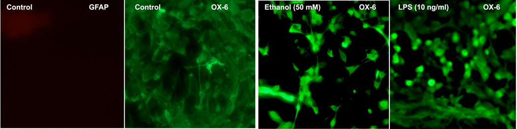
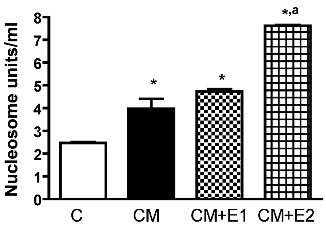
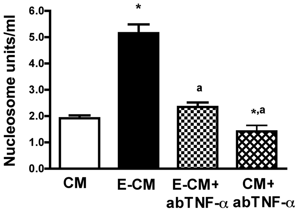
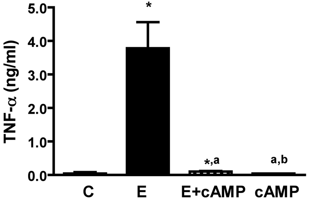
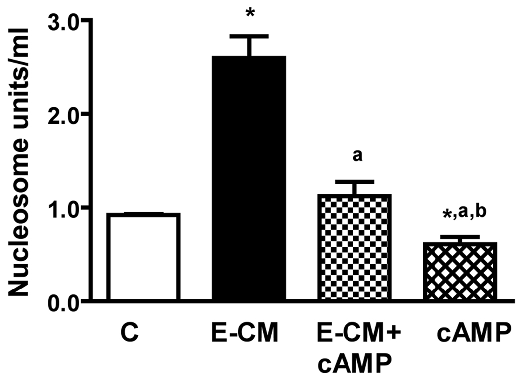
Methods: Ethanol's neurotoxic action was determined on enriched fetal mediobasal hypothalamic neuronal cells with or without microglia cells or ethanol-activated microglia-conditioned media. Ethanol's apoptotic action was determined using nucleosome assay. Microglia activation was determined using OX6 histochemistry and by measuring inflammatory cytokines secretion from microglia in cultures using enzyme-linked immunosorbent assay (ELISA). An immunoneutralization study was conducted to identify the role of a cytokine involved in ethanol's apoptotic action.

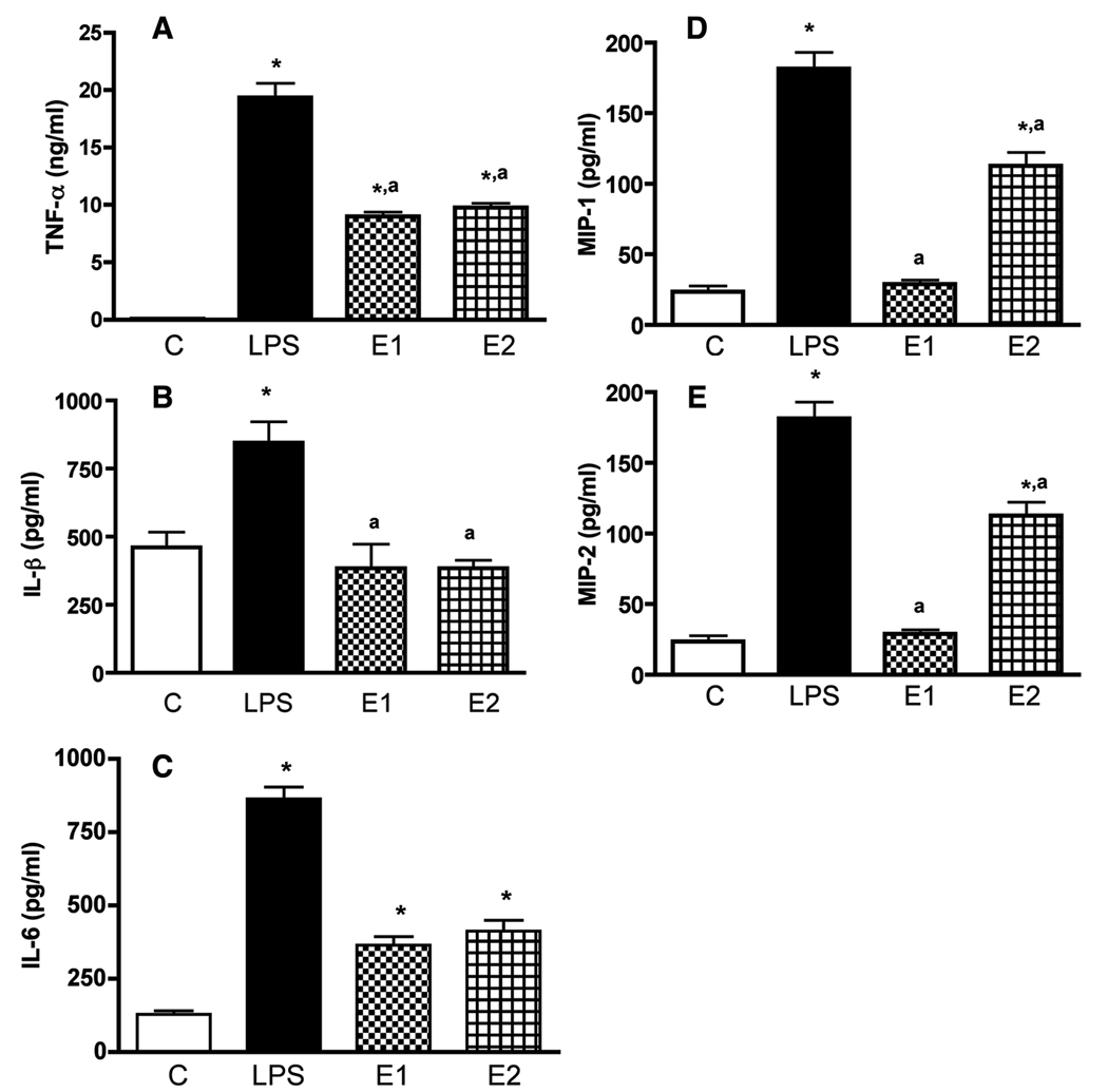
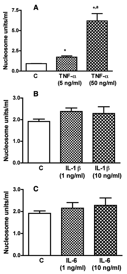
Results: We show here that ethanol at a dose range of 50 and 100 mM induces neuronal death by an apoptotic process. Ethanol's ability to induce an apoptotic death of neurons is increased by the presence of ethanol-activated microglia-conditioned media. In the presence of ethanol, microglia showed elevated secretion of various inflammatory cytokines, of which TNF-α shows significant apoptotic action on mediobasal hypothalamic neuronal cells. Ethanol's neurotoxic action was completely prevented by cAMP. The cell-signaling molecule also prevented ethanol-activated microglial production of TNF-α. Immunoneutralization of TNF-α prevented the microglia-derived media's ability to induce neuronal death.

Conclusions: These results suggest that ethanol's apoptotic action on hypothalamic neuronal cells might be mediated via microglia, possibly via increased production of TNF-α. Furthermore, cAMP reduces TNF-α production from microglia to prevent ethanol's neurotoxic action.


Figures

Fig.1
Fig.1. The effect of ethanol on microglia activation: histochemical evidence
Microglial cells were isolated and cultured on slide chambers for 3 days. The cells were then exposed to control media (CONT) or media containing ethanol 50 mM or LPS (10 ng/ml) for 24 h. Cells were then processed and stained for activated microglia marker (OX-6) or the astrocyte marker glial fibrillary acidic protein (GFAP).
Fig.2
Fig.2. The effect of ethanol and LPS on TNF-α, IL-1β, IL-6, MIP-1α and MIP-2 secretion from cultured microglia in primary cultures
Microglial cells were isolated and cultured in 24 well plates (1×10⁵) and treated for 24 h with 50 mM ethanol (E1) or 100 mM (E2). A group of cultures was treated with 10 ng/ml lipopolyssacaride (LPS) as a positive control. Control cultures were treated with vehicle only (C). The media samples were collected and used for the measurement of TNF-α (A), IL-1β (B), IL-6 (C), MIP-1α (D) and MIP-2 (E) by ELISA. Each bar represents mean ± SEM of 4–6 samples. *P< 0.001, as compared to C. aP< 0.001, as compared to LPS.
Fig.3
Fig.3. The effect of ethanol-activated microglia conditioned media on an apoptotic cell death of fetal MBH neurons
Fetal MBH neuronal cells were isolated and cultured in T25 flasks (1×10⁶/well) for 2 days. The cells were then exposed for 24 h with vehicle (C) or conditioned medium from microglia cultures (1×10⁵ cells) treated for 24 h with no ethanol (CM), 50 mM ethanol (CM + E1), or 100 mM (CM + E2). The neuron culture cell lysates were used to determine apoptotic death using a nucleosome ELISA. Each bar represents mean ± SEM of 5–6 samples. *P< 0.001, as compared to C. aP< 0.001, as compared to CM.
Fig.4
Fig.4. Effects of TNF-α IL-1β and IL-6 on apoptosis of MBH neurons
Fetal MBH neuronal cells were isolated and cultured in T25 flasks (1×10⁶/well) for 2 days. The cells were then exposed for 24 h with various doses of TNF-α (A), IL-1β (B) or IL-6 (C) for a period of 24 h. Cell lysates were used to determine apoptotic death using a nucleosome ELISA. Each bar represents mean ± SEM of 5–6 simples. *P< 0.001, as compared to C. aP< 0.001, as compared to TNF-α (5 ng/ml).
Fig.5
Fig.5. The effect of immunoneutralization of TNF-α on the ability of ethanol-activated microglia conditioned media to induce apoptosis of MBH neurons
Fetal MBH neuronal cells were isolated and cultured in T25 flasks (1×10⁶/well) for 2 days. The cells were then exposed to conditioned medium from microglia treated for 24 h with 50 mM ethanol (E-CM) or no ethanol (CM). A dose of 10 µg/ml antibody to TNF-α was then added to E-CM (E-CM + ab-TNF-α) or CM (CM + ab-TNF-α) mixed and added to neuron cultures to determine neuronal apoptosis by nucleosome ELISA. Each bar represents mean ± SEM of 5–6 samples. *P< 0.001, as compared to CM. aP< 0.001, as compared to E-CM.
Fig.6
Fig.6. Effects of dbcAMP on basal and ethanol-induced release of TNF-α from microglial cells
Microglial cells were isolated and cultured in 24 well plates (1×10⁵) and treated for 24 h with a 50 mM dose of ethanol without (E) or with a 1µM/ml dose of dbcAMP (E + cAMP), or with a 1µM/ml dose of dbcAMP alone (cAMP) for a period of 24 h. Control cultures were treated with vehicle only (C). The media samples were collected and used for the measurement of TNF-α by ELISA. Each bar represents mean ± SEM of 5–6 samples. *P< 0.001, as compared to C. aP< 0.001, as compared to E-CM. bP< 0.001, as compared to E-CM + cAMP.
Fig.7
Fig.7. Effects of dbcAMP on the ability of ethanol-activated microglia conditioned media to induce apoptosis of MBH neurons
Microglial cells were isolated and cultured in 24 well plates (1×10⁵) and treated for 24 h with a 50 mM dose of ethanol without (E) or with a 1µM/ml dose of dbcAMP (E + cAMP), or with a 1µM/ml dose of dbcAMP alone (cAMP) for a period of 24 h. Control cultures were treated with vehicle only (C). Cell lysates were used to determine apoptotic death using a nucleosome ELISA. Each bar represents mean ± SEM of 5–6 samples. *P< 0.01, as compared to C. aP< 0.001, as compared to E-CM. bP< 0.001, as compared to E-CM+cAMP.

Similar articles

Cited by

References

    1. Allen RT, Hunter WJ, III, Agrawal WJDK. Morphological and biological characterization and analysis of apoptosis. J Pharmacol Toxicol Methods. 1997;37:215–228. 3rd. - PubMed
    1. Bellucci A, Westwood AJ, Ingram E, Casamenti F, Goedert M, Spillantini MG. Induction of inflammatory mediators and microglial activation in mice transgenic for mutant human P301S tau protein. Am J Pathol. 2004;165:1643–1652. - PMC - PubMed
    1. Boyadjieva N, Sarkar DK. Effects of ethanol on basal and prostaglandin E1-induced increases in beta-endorphin release and intracellular cAMP levels in hypothalamic cells. Alcoholism: Clin Exp Res. 1997;21:1005–1009. - PubMed
    1. Boyadjieva N, Sarkar DK. Effects of ethanol on basal and adenosine-induced increases in beta-endorphin release and intracellular cAMP levels in hypothalamic cells. Brain Res. 1999;824:112–118. - PubMed
    1. Brown RE. An Introduction to Neuroendocrinology. New York, NY: Cambridge University Press; 1998. pp. 41–43.

Publication types

MeSH terms