

Stromal cells in the human gut show ultrastructural features of fibroblasts and smooth muscle cells but not myofibroblasts
- PMID: 20662992
- PMCID: PMC3823193
- DOI: 10.1111/j.1582-4934.2010.01132.x
Stromal cells in the human gut show ultrastructural features of fibroblasts and smooth muscle cells but not myofibroblasts
Abstract
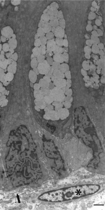
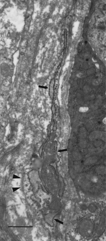
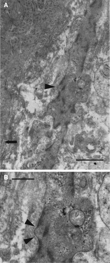
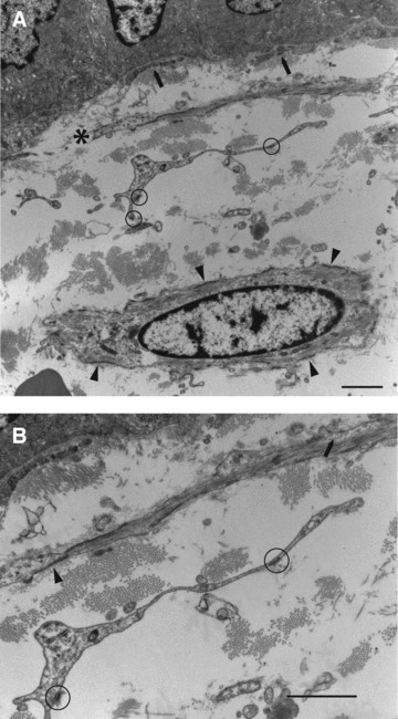
The free spindled cells of the lamina propria of the gut have been reported as showing fibroblastic, smooth-muscle and myofibroblastic differentiation. A precise understanding of the differentiation of these cells is essential for appreciating their functions, and this paper addresses this question using ultrastructural analysis. Histologically normal samples from different areas of the gastrointestinal tract were studied. Both subepithelial stromal cells, lying immediately beneath the basal lamina, and the deeper interstitial stromal cells, were studied. Subepithelial and interstitial cells had comparable features, reinforcing the idea that these formed a single reticulum of cells. Two major cell types were identified. Some were smooth-muscle cells, on the basis of abundant myofilaments with focal densities, glycogen, an irregular cell surface, focal lamina and multiple attachment plaques alternating with plasmalemmal caveolae. Some cells had a lesser expression of these markers, especially of myofilaments, and were regarded as poorly differentiated smooth-muscle cells and descriptively referred to as 'myoid'. Other cells were fibroblastic to judge by prominent rough endoplasmic reticulum, an absence of myofilaments and lamina, but presence of focal adhesions. The fibronexus junctions of true myofibroblasts were not seen. The study emphasises that the smooth-muscle actin immunoreactivity in this anatomical site resides in smooth-muscle cells and not in myofibroblasts, a view consistent with earlier ultrastructural and immunostaining results. The recognition that these cells are showing smooth-muscle or fibroblastic but not true myofibroblastic differentiation should inform our understanding of the function of these cells.
© 2011 The Authors Journal of Cellular and Molecular Medicine © 2011 Foundation for Cellular and Molecular Medicine/Blackwell Publishing Ltd.
Figures


Similar articles
-
The fibronexus in reactive and tumoral myofibroblasts: further characterisation by electron microscopy.Histol Histopathol. 2001 Jan;16(1):57-70. doi: 10.14670/HH-16.57. Histol Histopathol. 2001. PMID: 11193213
-
Electron microscopy in the study of myofibroblastic lesions.Semin Diagn Pathol. 2003 Feb;20(1):13-24. Semin Diagn Pathol. 2003. PMID: 12693672 Review.
-
Myofibroblastoma of breast: evidence favoring smooth-muscle rather than myofibroblastic differentiation.Ultrastruct Pathol. 1999 Jul-Aug;23(4):249-57. doi: 10.1080/019131299281581. Ultrastruct Pathol. 1999. PMID: 10503744
-
The myofibroblast: a study of normal, reactive and neoplastic tissues, with an emphasis on ultrastructure. Part 1--normal and reactive cells.J Submicrosc Cytol Pathol. 2005 Aug;37(2):109-204. J Submicrosc Cytol Pathol. 2005. PMID: 16335592 Review.
-
Actin filaments in human renal tubulo-interstitial fibrosis: significance for the concept of epithelial-myofibroblast transformation.J Submicrosc Cytol Pathol. 2003 Jul;35(3):221-33. J Submicrosc Cytol Pathol. 2003. PMID: 14690170
Cited by
-
Identification of telocytes in skeletal muscle interstitium: implication for muscle regeneration.J Cell Mol Med. 2011 Jun;15(6):1379-92. doi: 10.1111/j.1582-4934.2011.01330.x. J Cell Mol Med. 2011. PMID: 21609392 Free PMC article.
-
The Mesangial cell - the glomerular stromal cell.Nat Rev Nephrol. 2021 Dec;17(12):855-864. doi: 10.1038/s41581-021-00474-8. Epub 2021 Sep 10. Nat Rev Nephrol. 2021. PMID: 34508249 Review.
-
An integrated map of fibroblastic populations in human colon mucosa and cancer tissues.Commun Biol. 2022 Dec 3;5(1):1326. doi: 10.1038/s42003-022-04298-5. Commun Biol. 2022. PMID: 36463319 Free PMC article.
-
Telocytes and putative stem cells in the lungs: electron microscopy, electron tomography and laser scanning microscopy.Cell Tissue Res. 2011 Sep;345(3):391-403. doi: 10.1007/s00441-011-1229-z. Epub 2011 Aug 20. Cell Tissue Res. 2011. PMID: 21858462 Free PMC article.
-
The small and large intestine contain related mesenchymal subsets that derive from embryonic Gli1+ precursors.Nat Commun. 2023 Apr 21;14(1):2307. doi: 10.1038/s41467-023-37952-5. Nat Commun. 2023. PMID: 37085516 Free PMC article.
References
-
- Kaye GI, Lane N, Pascal RR. Colonic pericryptal fibroblast sheath: replication, migration, and cytodifferentiation of a mesenchymal cell system in adult tissue. II. Fine structural aspects of normal rabbit and human colon. Gastroenterology. 1968;54:852–65. - PubMed
-
- Marsh MN, Trier JS. Morphology and cell proliferation of subepithelial fibroblasts in adult mouse jejunum. I. Structural features. Gastroenterology. 1974;67:622–35. - PubMed
-
- Brenmoehl J, Falk W, Göke M, et al. Inflammation modulates fibronectin isoform expression in colonic lamina propria fibroblasts (CLPF) Int J Colorectal Dis. 2008;23:947–55. - PubMed
-
- Adegboyega PA, Mifflin RC, DiMari JF, et al. Immunohistochemical study of myofibroblasts in normal colonic mucosa, hyperplastic polyps, and adenomatous colorectal polyps. Arch Pathol Lab Med. 2002;126:829–36. - PubMed
-
- Güldner F-H, Wolff JR, Graf Keyserlingk D. Fibroblasts as a part of the contractile system in duodenal villi of rat. Z Zellforsch. 1972;135:349–60. - PubMed
MeSH terms
LinkOut - more resources
Full Text Sources
