. 2010 Jul 21:7:18.
doi: 10.1186/1743-8977-7-18.

Polycyclic aromatic hydrocarbon components contribute to the mitochondria-antiapoptotic effect of fine particulate matter on human bronchial epithelial cells via the aryl hydrocarbon receptor

Affiliations

Polycyclic aromatic hydrocarbon components contribute to the mitochondria-antiapoptotic effect of fine particulate matter on human bronchial epithelial cells via the aryl hydrocarbon receptor

Ioana Ferecatu et al. Part Fibre Toxicol. .

Abstract

Background: Nowadays, effects of fine particulate matter (PM2.5) are well-documented and related to oxidative stress and pro-inflammatory response. Nevertheless, epidemiological studies show that PM2.5 exposure is correlated with an increase of pulmonary cancers and the remodeling of the airway epithelium involving the regulation of cell death processes. Here, we investigated the components of Parisian PM2.5 involved in either the induction or the inhibition of cell death quantified by different parameters of apoptosis and delineated the mechanism underlying this effect.

Results: In this study, we showed that low levels of Parisian PM2.5 are not cytotoxic for three different cell lines and primary cultures of human bronchial epithelial cells. Conversely, a 4 hour-pretreatment with PM2.5 prevent mitochondria-driven apoptosis triggered by broad spectrum inducers (A23187, staurosporine and oligomycin) by reducing the mitochondrial transmembrane potential loss, the subsequent ROS production, phosphatidylserine externalization, plasma membrane permeabilization and typical morphological outcomes (cell size decrease, massive chromatin and nuclear condensation, formation of apoptotic bodies). The use of recombinant EGF and specific inhibitor led us to rule out the involvement of the classical EGFR signaling pathway as well as the proinflammatory cytokines secretion. Experiments performed with different compounds of PM2.5 suggest that endotoxins as well as carbon black do not participate to the antiapoptotic effect of PM2.5. Instead, the water-soluble fraction, washed particles and organic compounds such as polycyclic aromatic hydrocarbons (PAH) could mimic this antiapoptotic activity. Finally, the activation or silencing of the aryl hydrocarbon receptor (AhR) showed that it is involved into the molecular mechanism of the antiapoptotic effect of PM2.5 at the mitochondrial checkpoint of apoptosis.

Conclusions: The PM2.5-antiapoptotic effect in addition to the well-documented inflammatory response might explain the maintenance of a prolonged inflammation state induced after pollution exposure and might delay repair processes of injured tissues.


Figures

Figure 1
Figure 1
Effect of PM2.5-AW exposure on human bronchial epithelial cells. (A) Dose response of DiOC6(3) (to measure the mitochondrial ΔΨm drop), hydroethidine (HE, ROS-sensitive dye), Annexin V-FITC (for phosphatidylserine exposure) and the propidium iodide (PI, as a plasma membrane permeabilization marker) staining after 24 hours exposure of 16HBE bronchial epithelial cells to PM2.5-AW (1-50 μg/cm2) or H2O2 (500 μM). (B) Kinetic study of PM2.5-AW effect on apoptosis. 16HBE cells were exposed to PM2.5-AW (1 to 50 μg/cm2) or H2O2 (500 μM) for 48 h or 72 h before flow cytometric analysis of cells presenting simultaneously a mitochondrial depolarization (DiOC6(3) low) and a superoxide anion generation (HE -> Eth high). Results are representative of three independent experiments. (C) Subconfluent bronchial epithelial cell lines NCI-H292, BEAS-2B and primary bronchial epithelial cells (NHBE) were exposed 24 h to 1-50 μg/cm2 PM2.5-AW, and H2O2 (1 mM) before flow cytometric analysis of cells presenting simultaneously a DiOC6(3) low and PI high staining. (D) Human bronchial epithelial cells 16HBE, NCI-H292, BEAS-2B or NHBE were exposed 24 hours to 10 μg/cm2 of different batches of PM2.5 (Auteuil-Winter (AW), Auteuil-Summer (AS), Vitry-Winter (VW) or Vitry-Summer (VS) corresponding to two locations of Paris: (i) a school playground at Vitry-sur-Seine in a suburb of Paris and (ii) Porte d'Auteuil adjacent to a major highway). Then, cells were analyzed as previously described. Data are represented as mean ± SD (* treated vs. control, p < 0.05, n = 3).
Figure 2
Figure 2
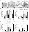
Parisian PM2.5 have an antiapoptotic effect against several death inducers. (A) Transmission electron microscopy of 16HBE cells treated 24 h with A23187 (3 μM) in the absence or in the presence of a 4 h pretreatment with 10 μg/cm2 PM2.5-AW. The micrographs illustrate that control or PM2.5-treated cells show normal nuclear chromatin condensation with the presence of some nucleoli (a) and normal ultrastructure of mitochondria (black arrow). PM's aggregates are localized near to the plasma membrane of cells exposed to particles or into the cytoplasm (white arrows). Treatment with A23187 alone triggered typical features of apoptotic cell death characterized by reduced cellular volume, massive chromatin condensation (b), formation of apoptotic bodies (black asterisk) and maintenance of the plasma membrane integrity. Note that PM2.5-AW pretreatment prevented A23187-induced morphological modifications and allowed 16HBE cells to retain nuclear or mitochondria morphologies similar to those of the control. Scale bar represent 5 μm. (B and C) After a 4 h pretreatment with PM2.5-AW (10 μg/cm2), 16HBE cells were exposed to different inducers of cell death for another 20 h such as mitochondrial respiratory chain inhibitors (rotenone (Rot, 5 μM), antimycin A (AMA, 25 μg/ml) and oligomycin (Omy, 5 μM)), calcium ionophores (ionomycin (Iono, 0.5 μM) and A23187, 3 μM), protein kinases inhibitor (staurosporine STS, 1 μM) and oxidative stress activator (H2O2, 500 μM). Then, cells were quantified for DiOC6(3) low or PI high staining by flow cytometry. (D and E) The human bronchial epithelial NCI-H292 cell line and NHBE primary cells were assessed by flow cytometry in the presence or the absence of PM2.5-AW pretreatement (10 μg/cm2) and apoptosis inducers: A23187 (3 μM), staurosporine (STS, 1 μM) and H2O2 (500 μM). Experiments were repeated three times, and means ± S.D. are shown. Significance was calculated with respect to untreated controls (*, p < 0.001) and with respect to non-pretreated cells with particles (#, p < 0.001).
Figure 3
Figure 3
Dose-effect studies of the antiapoptotic effect of PM2.5-AW. Apoptosis was quantified simultaneously by four parameters and expressed as percentage of 16HBE cells showing ΔΨm drop (DiOC6(3) low) and ROS production (HE->Eth high) or PS exposure (Annexin V+) and plasma membrane permeabilization (PI high). (A and B) After a 4 h pretreatment with PM2.5-AW (10 μg/cm2), 16HBE cells were exposed to the indicated concentrations of A23187. Significance was calculated with respect to medium conditions (*, p < 0.001, n = 3). (C and D) Particle pretreatment was performed with different doses (1-50 μg/cm2) 4 h prior to induction of apoptosis by 3 μM of A23187. Results are means ± S.D (n = 3). Significance was calculated with respect to non-pretreated cells with particles (#, p < 0.001).
Figure 4
Figure 4
Study of direct adsorption of A23187 onto particles. (a) 16HBE cells were exposed 4 h to 10 μg/cm2 PM2.5-AW prior to addition of 3 μM A23187 during 20 supplementary hours. (b) To avoid possible adsorption, PM2.5-AW were removed and cells were washed five times with PBS before drug treatment. (c) Particles and A23187 were co-incubated 4 h into a coated 24 wells-plate without cells, and then supernatant was used to trigger apoptosis for 20 h supplementary. ΔΨm dissipation was measured by flow cytometry and results are expressed as percentage of apoptosis induction as described in Materials and Methods section. Results are means ± S.D (n = 3). Significance was calculated for PM2.5-AW + A23187 versus A23187 alone for all conditions with p < 0.05 (*) or for PM2.5-AW + A23187 treatment with respect to a, b or c condition (#, p < 0.05).
Figure 5
Figure 5
The antiapoptotic effect is not correlated with proinflammatory cytokines release after PM2.5 exposure. (A) Proinflammatory potential of different batches of PM2.5 10 μg/cm2 (AW, AS, VW and VS) on 16HBE cells. After 4 h or 24 h particle exposure, Amphiregulin (AR) and granulocyte monocyte colony-stimulating factor (GM-CSF) secretion were evaluated by ELISA. Data are represented as mean ± SD of triplicates. * indicates significance at p < 0.001, treated vs. control. (B) Flow cytometric analysis was done as before in the presence or absence of PM2.5-AW pretreatement and/or apoptosis inducer A23187 (3 μM). The implication of the EGFR was evaluated using the recombinant EGF ligand (rEGF 150 ng/ml) or the inhibitor AG1478 (1 μM). Data are represented as mean ± SD of three independent results (* treated vs. control p < 0.05; # vs. A23187 alone, p < 0.05).
Figure 6
Figure 6
Role of the different components of PM2.5 in the antiapoptotic effect. (A) 16HBE cells were treated 4 h with different batches of PM2.5 10 μg/cm2 (AW, AS, VW and VS), the equivalent concentration of organic extracts (Oex, 4.27 mg/ml), aqueous extracts (Aex), washed particles (wash, 10 μg/cm2) of PM2.5-AW and carbon black (CB, 10 μg/cm2) before a 20 h exposure to A23187 (3 μM). Apoptosis was assessed by flow cytometry and expressed as induction of apoptosis. Results are mean ± SD (n = 3). * p < 0.001 compared with A23187 alone. (B and C) Effect of heavy PAH on A23187-induced apoptosis. Treatments with PM2.5-AW (10 μg/cm2), the vehicle (Cylohexane, 1%), Benzo[a]pyrene (B(a)P, 270 nM), Dibenzo[a,h]anthracene (D,B(a,h)A, 35 nM), Benzo[g,h,i]perylene (B(g,h,i)P, 443 nM), Indeno[1,2,3-cd]pyrene (iP, 217 nM) and Benzo[b]fluoranthrene (B(b)F, 333 nM), were performed 4 h prior to induction of apoptosis by A23187 (3 μM). Results are mean ± SD (n = 4). * vs. control, p < 0.001; # vs. A23187, p < 0.05.
Figure 7
Figure 7
AhR pathway is involved in the antiapoptotic effect of Parisian PM2.5. (A) 16HBE epithelial cells were preincubated or not in the presence of the agonist beta-naphtoflavone (beta-NF, 20 μg/ml) or the antagonist alpha-naphtoflavone (alpha-NF, 10 μg/ml) one hour before the usual PM2.5-AW pretreatment and/or apoptosis inducer A23187 (3 μM). Apoptosis was measured by flow cytometry as above. Results are mean ± SD (n = 3). p < 0.001, * vs. Control, # vs. A23187, § vs. A23187 + PM2.5. (B) Effect of AhR silencing. (Upper panel) 16HBE cells were incubated during 48 h with AhR siRNA (AhR, 10 nM), a control siRNA (Co, 10 nM) or non-transfected (NT), then total cell extract (80 μg) were loaded onto gel and subjected to immunoblotting with anti-AhR and anti-Actin antibodies. Quantifications were performed as respect to Actin and illustrated as AhR/Actin ratio. (Lower Graph) 16HBE cells were incubated with siRNA for 48 h as above and then 4 h pretreated to PM2.5-AW before apoptosis induction with A23187 for 20 h supplementary. Results of flow cytometry (DiOC(6)3 low and IP high) are from transfected cells (Alexa Fluor 647 positive population) which were around 80% for all conditions, illustrated as mean ± SD (n = 4). p < 0.001, * vs non-treated siRNA Co; p < 0.001, # vs. A23187 siRNA Co; p < 0.005, § vs. A23187 + PM2.5 siRNA Co.
Figure 8
Figure 8
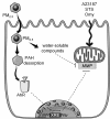
Hypothetic model for the mechanism of the antiapoptotic effect of PM2.5. Exposure of bronchial epithelial cells to PM2.5, leads to particles endocytosis and PAH desorption. Then, intracellular PAH can target and activate the aryl hydrocarbon receptor (AhR). It is noteworthy, that AhR translocates to the nucleus to bind to specific xenobiotic responsive elements (XRE) in the promoter of its target genes; some of them might be mitochondrial MMP regulators (dotted arrow), thus might protect the cell from apoptosis induced by A23187, staurosporin (STS) or oligomycin (Omy). Moreover, the water-soluble compounds of PM2.5 also have antiapoptotic activity, but the pathway involved is still under investigation (dotted arrow). Illustration carried out thanks to Servier Medical Art.

Similar articles

Cited by

References

    1. Boland S, Baeza-Squiban A, Marano F. Toxicité respiratoire des particules Diesel: les mécanismes cellulaires et moléculaires. Médecine Sciences. 2001;17:596–603.
    1. Baulig A, Poirault JJ, Ausset P, Schins R, Shi T, Baralle D, Dorlhene P, Meyer M, Lefevre R, Baeza-Squiban A, Marano F. Physicochemical characteristics and biological activities of seasonal atmospheric particulate matter sampling in two locations of Paris. Environ Sci Technol. 2004;38:5985–5992. doi: 10.1021/es049476z. - DOI - PubMed
    1. Baulig A, Garlatti M, Bonvallot V, Marchand A, Barouki R, Marano F, Baeza-Squiban A. Involvement of reactive oxygen species in the metabolic pathways triggered by diesel exhaust particles in human airway epithelial cells. Am J Physiol Lung Cell Mol Physiol. 2003;285:L671–679. - PubMed
    1. Boland S, Baeza-Squiban A, Fournier T, Houcine O, Gendron MC, Chevrier M, Jouvenot G, Coste A, Aubier M, Marano F. Diesel exhaust particles are taken up by human airway epithelial cells in vitro and alter cytokine production. Am J Physiol. 1999;276:L604–613. - PubMed
    1. Calcabrini A, Meschini S, Marra M, Falzano L, Colone M, De Berardis B, Paoletti L, Arancia G, Fiorentini C. Fine environmental particulate engenders alterations in human lung epithelial A549 cells. Environ Res. 2004;95:82–91. doi: 10.1016/j.envres.2003.07.011. - DOI - PubMed

MeSH terms