. 2010 Jul 21;3(1):60.
doi: 10.1186/1756-3305-3-60.

Hepatic stellate cells and parasite-induced liver fibrosis

Affiliations

Hepatic stellate cells and parasite-induced liver fibrosis

Barrie Anthony et al. Parasit Vectors. .

Abstract

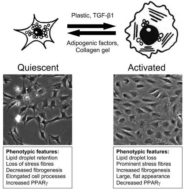
Fibrogenesis is a common feature of many diseases where there is severe insult to the liver. The hepatic stellate cell trans-differentiation into a myofibroblast has been identified as an important event in liver fibrogenesis and has been well investigated over the last few years in a number of liver diseases. The trans-differentiation process can be monitored in vitro by evaluation of biomarkers that are characteristic of normal quiescent hepatic stellate cells or activated myofibroblasts. Two major parasitic diseases associated with liver injury and fibrosis are schistosomiasis and echinococcosis. Recent studies have highlighted a role for activated hepatic stellate cells in both murine and human schistosomiasis as well as demonstrating that schistosome antigens are able to regulate this trans-differentiation process. Study of the hepatic stellate cell and its interaction with parasite-derived antigens may be pivotal in our understanding of the pathology associated with schistosomiasis and other parasitic diseases, including echinococcosis, as well as revealing new information on the trans-differentiation process in this cell type.


Figures

Figure 1
Figure 1
Summary of the process of hepatic stellate cell (HSC) trans-differentiation. The imaged cells are LX-2 cells taken after 72hrs growth with adipogenic factor induced quiescence on the left and activated cells on the right. The quiescent HSC phenotypically has a small cell body with cellular processes extending into the growth area around the cell. Lipid droplets are observed in the cells that are characterised by a lack of αSMA stress fibres, low expression of fibrosis associated genes (Col1A1, CTGF and αSMA) and increased expression of PPAR-γ. Upon activation, the cells lose their ability to store lipid droplets and have a large flat appearance. αSMA stress fibres are observed in the cells and gene expression of fibrosis associated genes are increased while PPAR-γ expression is reduced.

References

    1. Ross AG, Bartley PB, Sleigh AC, Olds GR, Li Y, Williams GM, McManus DP. Schistosomiasis. N Engl J Med. 2002;346:1212–1220. doi: 10.1056/NEJMra012396. - DOI - PubMed
    1. Vuitton DA. The ambiguous role of immunity in echinococcosis: protection of the host or of the parasite? Acta Trop. 2003;85:119–132. doi: 10.1016/S0001-706X(02)00230-9. - DOI - PubMed
    1. Leask A, Abraham DJ. TGF-beta signaling and the fibrotic response. FASEB J. 2004;18:816–827. doi: 10.1096/fj.03-1273rev. - DOI - PubMed
    1. Friedman SL. Liver fibrosis -- from bench to bedside. J Hepatol. 2003;38(Suppl 1):S38–53. doi: 10.1016/S0168-8278(02)00429-4. - DOI - PubMed
    1. Friedman SL. Mechanisms of hepatic fibrogenesis. Gastroenterology. 2008;134:1655–1669. doi: 10.1053/j.gastro.2008.03.003. - DOI - PMC - PubMed

LinkOut - more resources