. 2010 Jul 27:7:37.
doi: 10.1186/1476-9255-7-37.

Differential induction of inflammatory cytokines by dendritic cells treated with novel TLR-agonist and cytokine based cocktails: targeting dendritic cells in autoimmunity

Affiliations

Differential induction of inflammatory cytokines by dendritic cells treated with novel TLR-agonist and cytokine based cocktails: targeting dendritic cells in autoimmunity

Simon S Jensen et al. J Inflamm (Lond). .

Abstract

Background: Dendritic cells (DC) are main gate-keepers of the immune system, bridging the innate and adaptive immune system. DCs are able to mature into inflammatory DCs at sites of inflammation in both autoimmune and allergic disease, thereby sustaining a continuous activation of the adaptive immune system at sites of inflammation. This function of DCs makes them attractive target cells for therapeutic intervention in inflammatory diseases. We have designed a DC-based screening model by which drug candidates can be evaluated for their ability to suppress DC maturation into an inflammatory and disease promoting phenotype.

Methods: Human monocyte derived DCs were differentiated using IL-4 and GM-CSF to immature DCs (imDCs). The imDCs were treated with various combinations of TLR-agonists and pro-inflammatory cytokines to identify cocktails with ability to mature imDCs into inflammatory DCs. The effect of the cocktails on DC maturation was evaluated using ELISA and cytokine arrays to measure secreted cytokines and chemokines. FACS analysis was used to assess expression of maturation markers, and functional studies were carried out using naïve allogeneic T-cells to assay for a Th1-promoting DC phenotype.

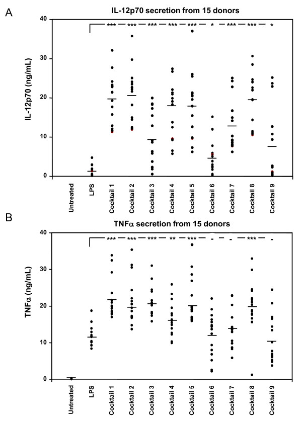
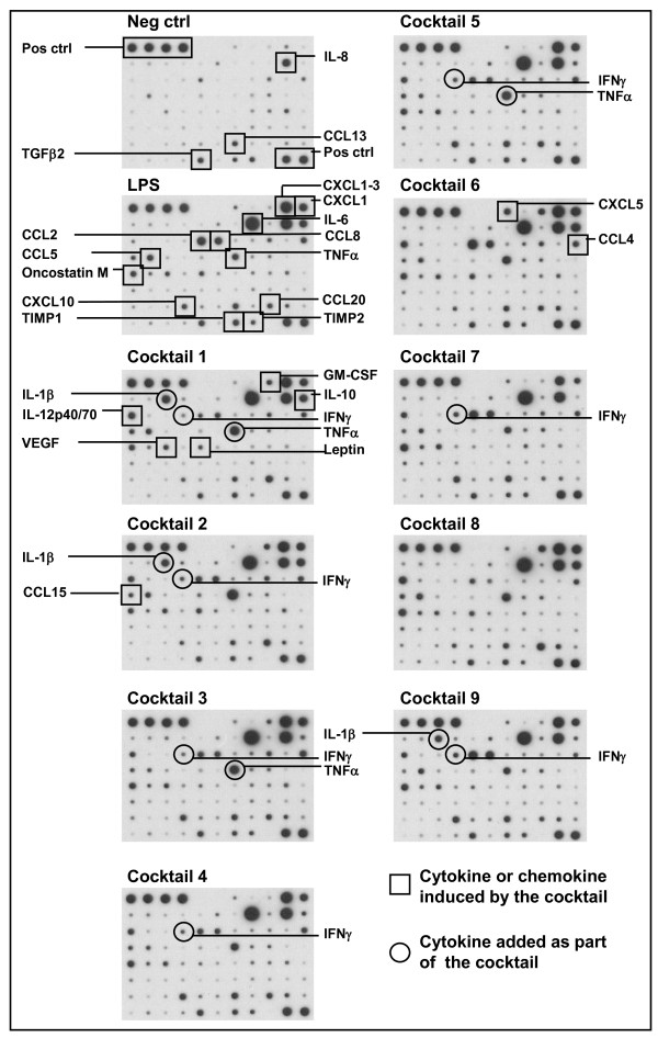
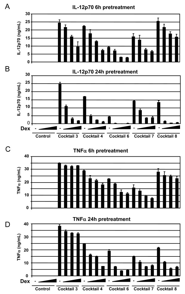
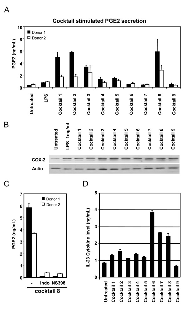
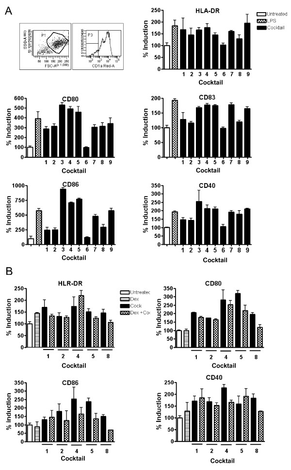
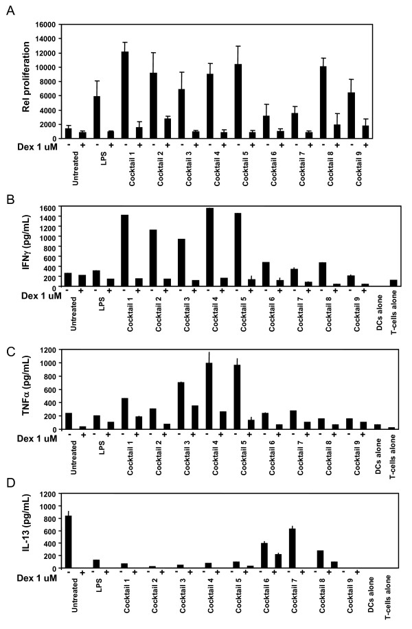
Results: Nine cocktails were designed with potent ability to induce secretion of the Th1-promoting cytokines IL-12p70 and TNFalpha from imDCs, and three were able to induce the Th17-promoting cytokine IL-23. The cocktails were further characterized using cytokine arrays, showing induction of inflammation related cytokines and chemokines like CXCL10, CCL2, CCL4, CCL8, CCL15, CCL20 and IL-8, of which some are present in a range of autoimmune pathologies. Prostaglandin E2 secretion was identified from DCs treated with TLR agonists poly I:C and peptidoglycan, but not LPS. The cocktails were able to induce DC maturation markers like HLA-DR, CD40, CD80, CD83 and CD86, except the TLR7/8 agonist R848. Functional end-points made by co-culture of allogeneic CD4+ T cells with the cocktail treated DCs, showed that five cocktails in particular could induce a classical Th1-phenotype with ability to secrete high amounts of the hall-mark cytokine IFNgamma. The model was validated using dexamethasone and two COX-inhibitors, which were able to suppress the cocktail driven pro-inflammatory DC maturation.

Conclusions: The identification of novel Th1-promoting cocktails allows screening of anti-inflammatory drug candidates by assessing the ability to suppress the activation and differentiation of imDCs into inflammatory DCs with a specific Th1-promoting phenotype. The model thus provides a screening tool, which can identify potential anti-inflammatory effects on the natural regulator of the immune response, the dendritic cell.


Figures

Figure 1
Figure 1
Cocktail screening and donor variation for IL-12p70 and TNFα secretion. A total of 15 different donor-derived imDCs were treated with LPS (0.1 μg/mL) or the 9 cocktails as indicated in table 1. After 24 h of incubation, IL-12p70 and TNFα levels in the conditioned media was determined by ELISA. A) The amount of IL-12p70 protein in each donor is indicated by a dot, and the average of all 15 donors indicated by a horizontal bar. B) Amount of TNFα protein was determined similarly in the 15 donors. Data were analyzed using unpaired, two sided t-test, (***P < 0.005, **P < 0.01, *P < 0.05).
Figure 2
Figure 2
Cytokine array using conditioned media from cocktail treated DCs. Cytokine array membranes were incubated with conditioned media from DCs treated with cocktails for 24 h. To compensate for donor variations the conditioned media from four donors was mixed. Upper left four spots and lower right two spots serves as positive controls on each membrane. On the figure constitutively secreted cytokines can be seen on the picture indicated Neg. ctrl, which were untreated cells. Each induced protein is marked only once, squares mark DC-produced cytokines and chemokines, and a circle marks a cytokine which is added as part of the cocktail (TNFα, IL-1β and IFNγ).
Figure 3
Figure 3
Dexamethasone prevents cocktail induction of IL-12p70 and TNFα. Immature DCs from a single donor shows a dexamethasone-mediated dose dependent suppression of IL-12p70 and TNFα secretion. Dexamethasone was pre-incubated with imDCs for 6 hours (A and C) or 24 hours (B and D) with increasing concentration of dexamethasone at 0-0.01-0.1 and 1.0 μM. Dexamethasone treatment without cocktail did not induce IL-12p70 or TNFα (first 4 bars). The cocktails used are indicated below each set of data, and their exact composition is seen in table 1. This shows one representative example out of three. Cell viability was not significantly affected by treatment with cocktail and/or dexamethasone (not shown).
Figure 4
Figure 4
Cocktail induced stimulation of inflammatory markers. Prostaglandin E2 was determined in conditioned media from two representative donor derived DC batches as described in M&M (A). PGE2 secretion was highest when imDCs were treated with cocktail 1, 2, 3 and 8 for 24 hours. B) COX2 protein levels were analysed by western blot of lysates from DCs treated with cocktails for 24 h. Actin was used as loading control, and results shown are representative blot from two donors. C) Inhibition of PGE2 secretion induced by cocktail 8 was shown by pre-incubation of imDCs from two different donors for 2 h with the unspecific COX inhibitor indomethazine (indo) at 10 μM or the specific COX-2 inhibitor NS398 at 10 μM. D) The ability of cocktails to induce secretion of IL-23, shown as an average of measurements on 3 different donors. Bars show standard deviation between the three donors.
Figure 5
Figure 5
FACS profile on cocktail stimulated DCs. Surface staining by flow cytometric analysis of immature (untreated), LPS and cocktail stimulated DCs. The surface expression of relevant activation markers was analyzed on day 7. A) A total of 5000 events were collected by gating hDCs defined by forward (FSC) and side-scatter (SSC) characteristics. All histograms were gated for CD1a cells (70-95%). All our cells were CD14 negative (data not shown). Flow cytomeric analysis of maturation markes were done for DCs from three donors stimulated with LPS or cocktails, and results normalized to the untreated DCs (average value of Mean florescence intensity for untreated DCs from the three donors was set to 100%). The vertical bars indicate standard deviation (SD) values. B) Phenotypic surface analysis of the suppressive effect of dex on cocktail treated human DCs from two donors. Pre-treatment of immature DCs with dex for 6 h before addition of selected cocktails reduced the expression of activation markers. Results for the different treatments have been normalized in proportion to the untreated DC. The vertical bars indicate (SD) values.
Figure 6
Figure 6
T-cell proliferation and secreted cytokines stimulated by cocktail treated DCs. MLR performed on CD4+ T cells and allogeneic DCs. Mature cocktail stimulated DCs were more potent inducers of T cell proliferation in the MLR than immature DCs. A pretreatment of DCs with dexamethasone for 6 h before addition of cocktails, significantly prevent CD4+ T cell proliferation to a level similar to immature DCs. The results are representative of three donors. A) CD4+ T cells were cultured with allogeneic DCs for 5 days, and mitomycin-C treated in order to inhibit their proliferation. Proliferation of CD4+ T cells was determined in the last 18-24 h of culture. Each column represents the mean cpm of four replicates. Vertical bars represent the SD. B) The amount of IFNγ production by T cells was measured in the supernatants after a restimulation with platebound anti-CD3 and soluble anti-CD28 mAbs for 24 h. In the conditioned media was also measured C) TNFα, and D) IL-13 by ELISA. Each column represents the mean of triplicate wells. Vertical bars represent the SD values.

Similar articles

Cited by

References

    1. Steinman RM, Banchereau J. Taking dendritic cells into medicine. Nature. 2007;449(27):419–426. doi: 10.1038/nature06175. - DOI - PubMed
    1. Shortman K, Shalin HN. Steady-state and inflammatory dendritic-cell development. Net Rev Immunol. 2007;7:19–30. doi: 10.1038/nri1996. - DOI - PubMed
    1. Roy CR, Mocarski ES. Pathogen subversion of cell-intrinsic innate immunity. Nat Immunol. 2007;8(11):1179–1187. doi: 10.1038/ni1528. - DOI - PubMed
    1. Bouma G, Strober W. The immunological and genetic basis of inflammatory bowel disease. Nat Rev Immunol. 2003;3:521–533. doi: 10.1038/nri1132. - DOI - PubMed
    1. Bachmann MF, Kopf M, Marsland BJ. Chemokines: more than just road signs. Nat Rev Immunol. 2006;6:159–164. doi: 10.1038/nri1776. - DOI - PubMed