

ImmunoRatio: a publicly available web application for quantitative image analysis of estrogen receptor (ER), progesterone receptor (PR), and Ki-67
- PMID: 20663194
- PMCID: PMC2949645
- DOI: 10.1186/bcr2615
ImmunoRatio: a publicly available web application for quantitative image analysis of estrogen receptor (ER), progesterone receptor (PR), and Ki-67
Abstract
Introduction: Accurate assessment of estrogen receptor (ER), progesterone receptor (PR), and Ki-67 is essential in the histopathologic diagnostics of breast cancer. Commercially available image analysis systems are usually bundled with dedicated analysis hardware and, to our knowledge, no easily installable, free software for immunostained slide scoring has been described. In this study, we describe a free, Internet-based web application for quantitative image analysis of ER, PR, and Ki-67 immunohistochemistry in breast cancer tissue sections.
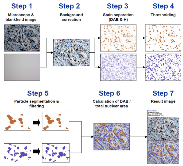
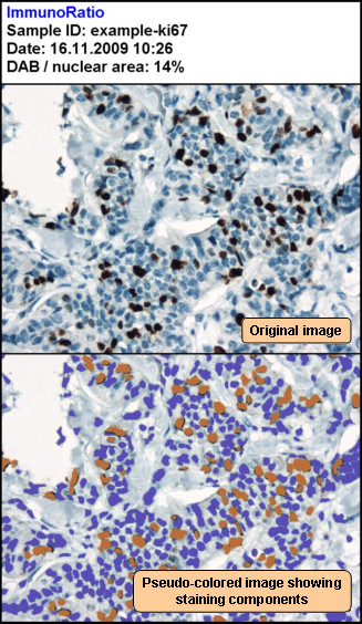
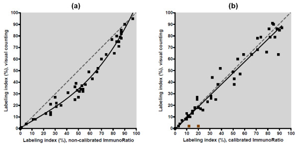
Methods: The application, named ImmunoRatio, calculates the percentage of positively stained nuclear area (labeling index) by using a color deconvolution algorithm for separating the staining components (diaminobenzidine and hematoxylin) and adaptive thresholding for nuclear area segmentation. ImmunoRatio was calibrated using cell counts defined visually as the gold standard (training set, n = 50). Validation was done using a separate set of 50 ER, PR, and Ki-67 stained slides (test set, n = 50). In addition, Ki-67 labeling indexes determined by ImmunoRatio were studied for their prognostic value in a retrospective cohort of 123 breast cancer patients.
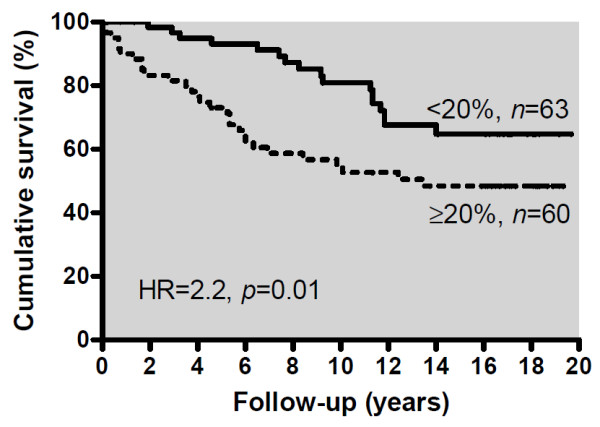
Results: The labeling indexes by calibrated ImmunoRatio analyses correlated well with those defined visually in the test set (correlation coefficient r = 0.98). Using the median Ki-67 labeling index (20%) as a cutoff, a hazard ratio of 2.2 was obtained in the survival analysis (n = 123, P = 0.01). ImmunoRatio was shown to adapt to various staining protocols, microscope setups, digital camera models, and image acquisition settings. The application can be used directly with web browsers running on modern operating systems (e.g., Microsoft Windows, Linux distributions, and Mac OS). No software downloads or installations are required. ImmunoRatio is open source software, and the web application is publicly accessible on our website.
Conclusions: We anticipate that free web applications, such as ImmunoRatio, will make the quantitative image analysis of ER, PR, and Ki-67 easy and straightforward in the diagnostic assessment of breast cancer specimens.
Figures
Similar articles
-
Clinical usefulness of the free web-based image analysis application ImmunoRatio for assessment of Ki-67 labelling index in breast cancer.J Clin Pathol. 2017 Aug;70(8):715-719. doi: 10.1136/jclinpath-2016-204162. Epub 2017 Mar 15. J Clin Pathol. 2017. PMID: 28298390 Free PMC article.
-
Technical note on the validation of a semi-automated image analysis software application for estrogen and progesterone receptor detection in breast cancer.Diagn Pathol. 2011 Jan 18;6:6. doi: 10.1186/1746-1596-6-6. Diagn Pathol. 2011. PMID: 21244664 Free PMC article.
-
Comparative analysis of methods for accurate recognition of cells through nuclei staining of Ki-67 in neuroblastoma and estrogen/progesterone status staining in breast cancer.Anal Quant Cytol Histol. 2009 Feb;31(1):49-62. Anal Quant Cytol Histol. 2009. PMID: 19320193
-
Novel image analysis approach for quantifying expression of nuclear proteins assessed by immunohistochemistry: application to measurement of oestrogen and progesterone receptor levels in breast cancer.Breast Cancer Res. 2008;10(5):R89. doi: 10.1186/bcr2187. Epub 2008 Oct 23. Breast Cancer Res. 2008. PMID: 18947395 Free PMC article.
-
An optimized image analysis algorithm for detecting nuclear signals in digital whole slides for histopathology.Cytometry A. 2017 Jun;91(6):595-608. doi: 10.1002/cyto.a.23124. Epub 2017 May 4. Cytometry A. 2017. PMID: 28472544
Cited by
-
Aptamer-Assisted Detection of the Altered Expression of Estrogen Receptor Alpha in Human Breast Cancer.PLoS One. 2016 Apr 4;11(4):e0153001. doi: 10.1371/journal.pone.0153001. eCollection 2016. PLoS One. 2016. PMID: 27043307 Free PMC article.
-
Methylselenocysteine preventing castration-resistant progression of prostate cancer.Prostate. 2015 Jun 15;75(9):1001-8. doi: 10.1002/pros.22987. Epub 2015 Mar 8. Prostate. 2015. PMID: 25754033 Free PMC article.
-
Exploring the spatial dimension of estrogen and progesterone signaling: detection of nuclear labeling in lobular epithelial cells in normal mammary glands adjacent to breast cancer.Diagn Pathol. 2014;9 Suppl 1(Suppl 1):S11. doi: 10.1186/1746-1596-9-S1-S11. Epub 2014 Dec 19. Diagn Pathol. 2014. PMID: 25565114 Free PMC article.
-
An enhanced chemopreventive effect of methyl donor S-adenosylmethionine in combination with 25-hydroxyvitamin D in blocking mammary tumor growth and metastasis.Bone Res. 2020 Jul 22;8:28. doi: 10.1038/s41413-020-0103-6. eCollection 2020. Bone Res. 2020. PMID: 32714613 Free PMC article.
-
uPAR antibody (huATN-658) and Zometa reduce breast cancer growth and skeletal lesions.Bone Res. 2020 Apr 17;8:18. doi: 10.1038/s41413-020-0094-3. eCollection 2020. Bone Res. 2020. PMID: 32337090 Free PMC article.
References
-
- Allred DC, Carlson RW, Berry DA, Burstein HJ, Edge SB, Goldstein LJ, Gown A, Hammond ME, Iglehart JD, Moench S, Pierce LJ, Ravdin P, Schnitt SJ, Wolff AC. NCCN Task Force Report: Estrogen Receptor and Progesterone Receptor Testing in Breast Cancer by Immunohistochemistry. J Natl Compr Canc Netw. 2009;7:S1–S21. quiz S22-23. - PubMed
-
- United Kingdom National External Quality Assessment Service (UK NEQAS) http://www.ukneqas.org.uk/ - PubMed
-
- Nordic immunohistochemical Quality Control (NordiQC) http://www.nordiqc.org/
-
- de Azambuja E, Cardoso F, de Castro G Jr, Colozza M, Mano MS, Durbecq V, Sotiriou C, Larsimont D, Piccart-Gebhart MJ, Paesmans M. Ki-67 as prognostic marker in early breast cancer: a meta-analysis of published studies involving 12,155 patients. Br J Cancer. 2007;96:1504–1513. doi: 10.1038/sj.bjc.6603756. - DOI - PMC - PubMed
Publication types
MeSH terms
Substances
LinkOut - more resources
Full Text Sources
Other Literature Sources
Research Materials
