

Trifocal monomyelomeric spinal cord arteriovenous fistulae in a seven-year-old boy
- PMID: 20663337
- PMCID: PMC3621531
- DOI: 10.1177/159101990100700205
Trifocal monomyelomeric spinal cord arteriovenous fistulae in a seven-year-old boy
Abstract
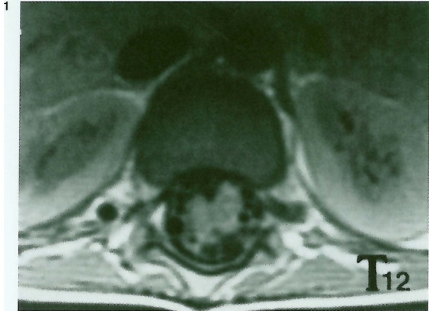
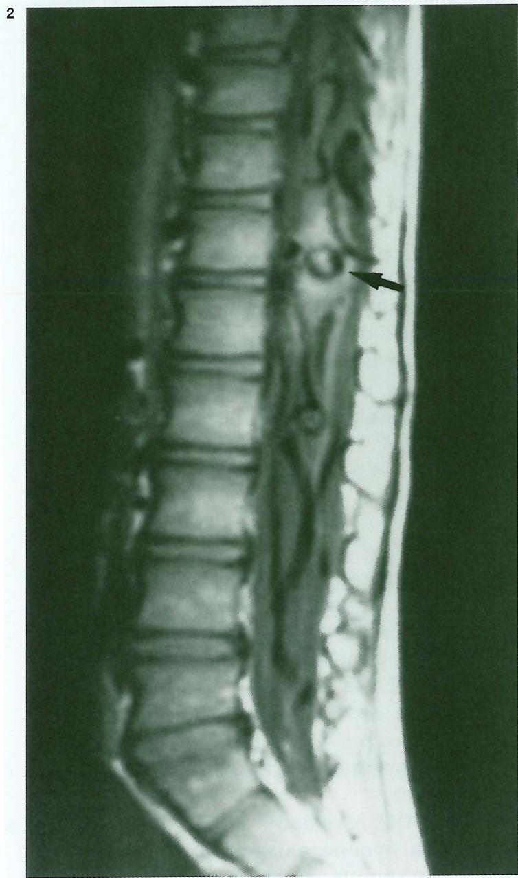
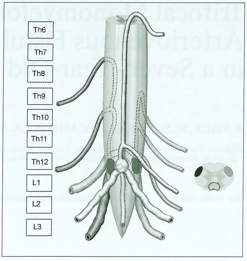
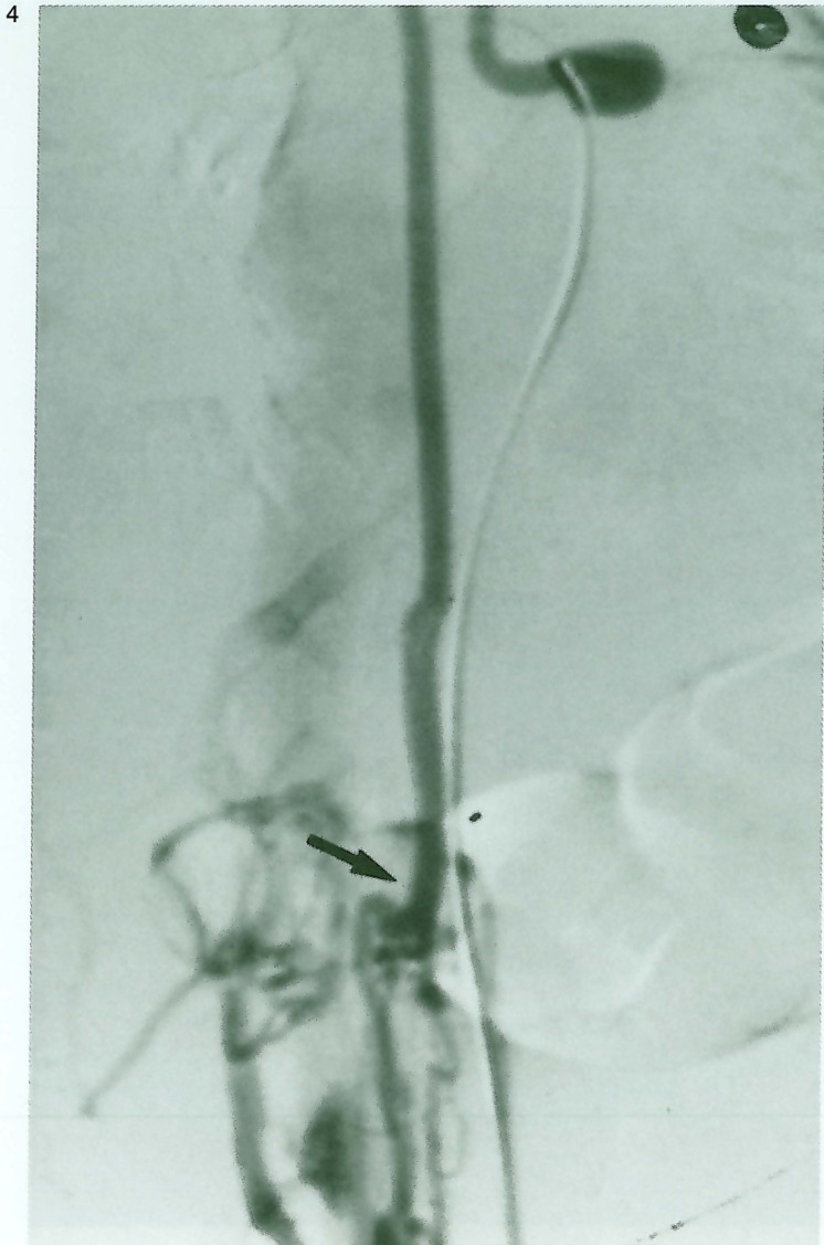
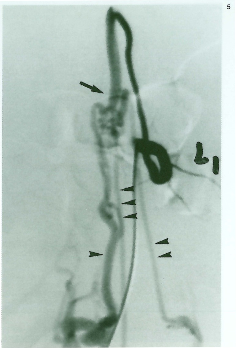
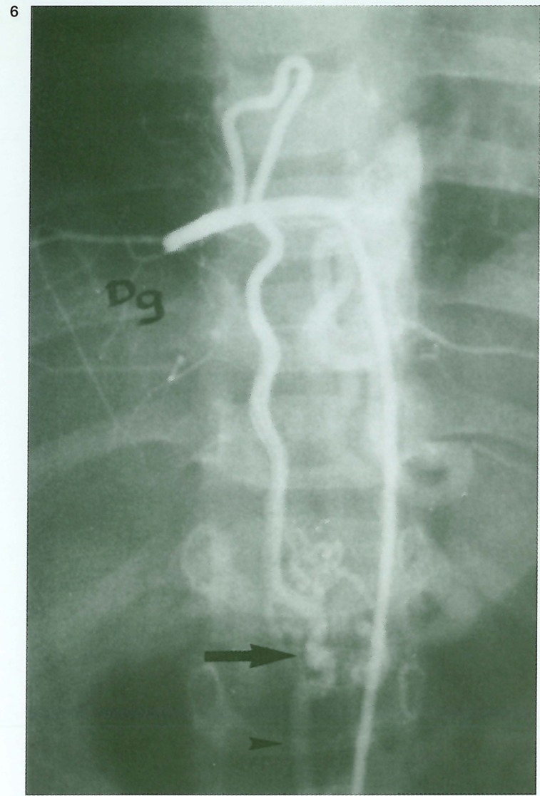
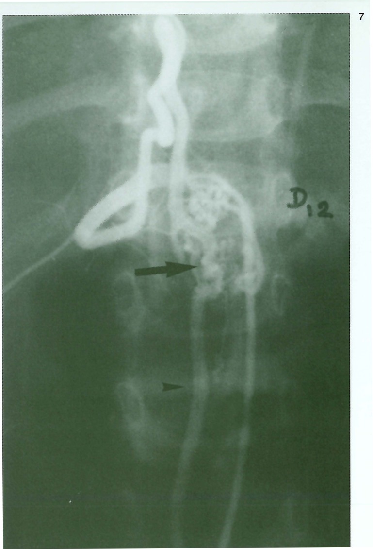
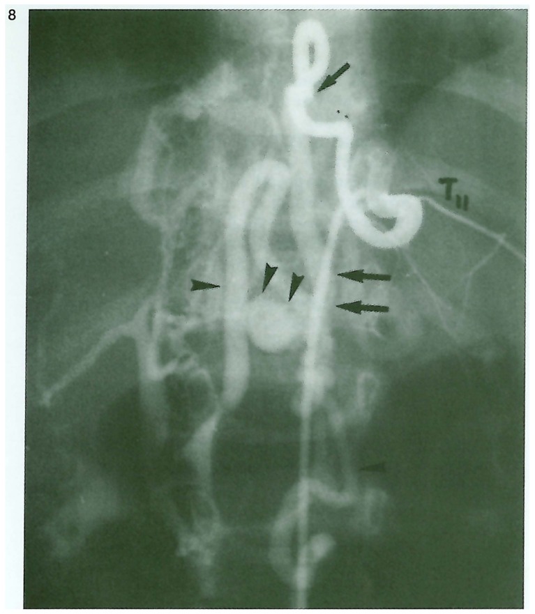
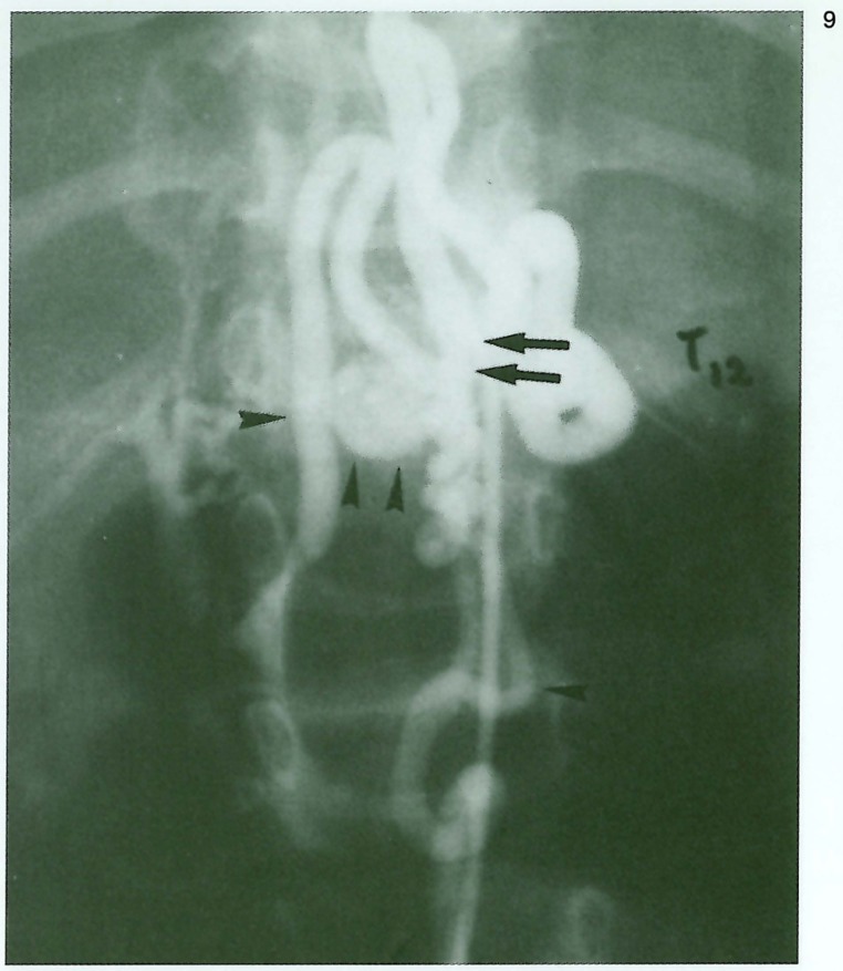
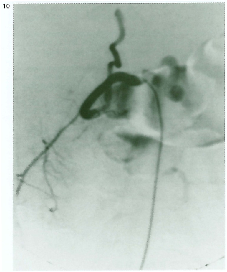
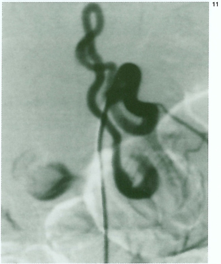
We describe a rare case of multiple arteriovenous fistulae of the spinal cord (SCAVF) in the same myelomer in a five-year-old boy. This case report consists of a trifocal SCAVF at the Th12 myelomeric level without communication between the three different fistulae. Two AVF were located posteriorly, bilateraly, in the spinal cord, fed by left and right posterior radiculopial arteries and one anteriorly in the anterior spinal axis. The venous drainage was independent for each lesion. The patient presents associated lesions characterized by cutaneous stain and inferior limb asymmetry. A metameric distribution is the explanation for the multiplicity of these lesions in a syndromic association related to Cobb syndrome. The patient was treated by transarterial embolization using glue with occlusion of the three different fistulae. The patient achieved a good improvement in neurological status.
Figures

Similar articles
-
Multifocal and metameric spinal cord arteriovenous malformations. Review of 19 cases.Interv Neuroradiol. 1999 Mar 30;5(1):27-34. doi: 10.1177/159101999900500105. Epub 2001 May 15. Interv Neuroradiol. 1999. PMID: 20670488 Free PMC article.
-
Spinal arteriovenous malformation associated with a radicular arteriovenous fistula suggested a metameric disease. A case report.Interv Neuroradiol. 2003 Mar 30;9(1):75-8. doi: 10.1177/159101990300900113. Epub 2004 Oct 22. Interv Neuroradiol. 2003. PMID: 20591307 Free PMC article.
-
Intraoperative direct puncture and embolization (IOPE) using a glue material for spinal cord arteriovenous fistula: a case report.Eur Spine J. 2015 May;24 Suppl 4:S594-9. doi: 10.1007/s00586-015-3773-9. Epub 2015 Feb 1. Eur Spine J. 2015. PMID: 25638046
-
[Intracranial dural fistula with spinal cord venous drainage. Apropos of 2 cases].J Neuroradiol. 1994 Apr;21(2):134-54. J Neuroradiol. 1994. PMID: 8014658 Review. French.
-
Exclusively epidural spinal metameric arteriovenous shunts: case report and literature review.Spine J. 2015 Mar 1;15(3):e15-22. doi: 10.1016/j.spinee.2014.11.022. Epub 2014 Nov 29. Spine J. 2015. PMID: 25450654 Review.
References
-
- Cogen P, Stein B. Spinal arteriovenous malformations with significant intramedullary components. J Neurosurg. 1983;59:471–487. - PubMed
-
- Meisel HJ, Lasjaunias P, Brock M. Interventional Neuroradiology. Vol. 38. Springer-Verlag; 1996. Multiple arteriovenous malformations of the spinal cord in a adolescent. Case report; pp. 490–493. - PubMed
-
- Garcia-Monaco, et al. Neuroradiology. Vol. 37. Springer-Verlag; 1995. Pial AVF in children as presenting manifestation of ROW disease. Interventional Neuroradiology; pp. 60–64. - PubMed
-
- Djindjian M, et al. Intradural extramedullary spinal arteriovenous malformations fed by the anterior spinal artery. Surg Neurol. 1977;8:85–93. - PubMed
-
- Rodesch, et al. Spinal cord AVM in a pediatric population. Children below 15 years of age. The place of Endovascular Management. Interventional Neuroradiology. 1995;1:29–42. - PubMed
LinkOut - more resources
Full Text Sources
