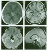
Spontaneous healing and complete disappearance of a giant basilar tip aneurysm in a child
- PMID: 20663341
- PMCID: PMC3621535
- DOI: 10.1177/159101990100700209
Spontaneous healing and complete disappearance of a giant basilar tip aneurysm in a child
Abstract
We report a rare case of spontaneous total thrombosis of a giant basilar tip aneurysm resulting in compression of the brainstem, diagnosed in a two-year-old child who presented with neurological deficits and third cranial nerve impairment. After conservative treatment, the giant aneurysm was completely thrombosed and the clinical symptoms were remarkably improved. MRI demonstrated dramatic shrinkage and ultimately complete disappearance of the giant aneurysm at seven month follow-up.
Figures


Similar articles
-
Guglielmi detachable coil treatment of a partially thrombosed giant basilar artery aneurysm in a child.Neuroradiology. 2000 Feb;42(2):142-4. doi: 10.1007/s002340050034. Neuroradiology. 2000. PMID: 10663494
-
[Giant partially thrombosed aneurysm of the vertebral artery: a case report and literature review].Zh Vopr Neirokhir Im N N Burdenko. 2016;80(5):106-115. doi: 10.17116/neiro2016805106-115. Zh Vopr Neirokhir Im N N Burdenko. 2016. PMID: 28635695 Review. Russian.
-
Successful shrinkage of a recurrent partially thrombosed symptomatic large basilar tip aneurysm using a Target 3D Coil.Surg Neurol Int. 2024 Mar 22;15:103. doi: 10.25259/SNI_44_2024. eCollection 2024. Surg Neurol Int. 2024. PMID: 38628531 Free PMC article.
-
Neurological Deterioration after Decompressive Suboccipital Craniectomy in a Patient with a Brainstem-compressing Thrombosed Giant Aneurysm of the Vertebral Artery.J Cerebrovasc Endovasc Neurosurg. 2016 Jun;18(2):115-119. doi: 10.7461/jcen.2016.18.2.115. Epub 2016 Jun 30. J Cerebrovasc Endovasc Neurosurg. 2016. PMID: 27790402 Free PMC article.
-
Thromboembolic complication induced stable occlusion of a ruptured basilar tip aneurysm. Case report and review of the literature.Interv Neuroradiol. 2010 Mar;16(1):83-8. doi: 10.1177/159101991001600111. Epub 2010 Mar 25. Interv Neuroradiol. 2010. PMID: 20377984 Free PMC article. Review.
Cited by
-
Transformation of a cranial fusiform aneurysm into a pseudotumoral-like mass prior to spontaneous occlusion and regression.Interv Neuroradiol. 2011 Mar;17(1):70-3. doi: 10.1177/159101991101700111. Epub 2011 Apr 29. Interv Neuroradiol. 2011. PMID: 21561561 Free PMC article.
-
Repair process in spontaneous intradural dissecting aneurysms in children: report of eight patients and review of the literature.Childs Nerv Syst. 2009 Jan;25(1):55-62. doi: 10.1007/s00381-008-0698-1. Epub 2008 Aug 19. Childs Nerv Syst. 2009. PMID: 18712397 Review.
-
Intracranial aneurysms in children aged under 15 years: review of 59 consecutive children with 75 aneurysms.Childs Nerv Syst. 2005 Jun;21(6):437-50. doi: 10.1007/s00381-004-1125-x. Epub 2005 Apr 15. Childs Nerv Syst. 2005. PMID: 15834727 Review.
-
Morphologic Change of Flow-Related Aneurysms in Brain Arteriovenous Malformations after Stereotactic Radiosurgery.AJNR Am J Neuroradiol. 2019 Apr;40(4):675-680. doi: 10.3174/ajnr.A6018. AJNR Am J Neuroradiol. 2019. PMID: 30948381 Free PMC article.
References
-
- Pia HW, Zierski J, et al. Giant Cerebral Aneurysm. In: Pia HW, editor. Cerebral Aneurysm. Heidelbterg: Spring Verlag; 1979. p. 336.
-
- Laughlin S, Terbrugge KG, et al. Endovascular management of paediatric intracranial aneurysms. Interventional Neuroradiology. 1997;3:205–214. - PubMed
-
- First MM, Cekirge S, et al. Guglielmi detachable coil treatment of a partially thrombosed giant basilar artery aneurysm in a child. Neuroradiology. 2000;42:142–144. - PubMed
-
- Atkinson JLD, Lane JI, et al. Spontaneous thrombosis of posterior cerebral artery aneurysm with angiographic reappearance. J Neurosurg. 1993;79:434–437. - PubMed
-
- Fukuoka S, Suematsu K, et al. Completely thrombosed giant aneurysm of the angular artery. Sur Neurol. 1984;22:145–148. - PubMed
LinkOut - more resources
Full Text Sources

