

Molecular diversity in ductal carcinoma in situ (DCIS) and early invasive breast cancer
- PMID: 20663721
- PMCID: PMC5527914
- DOI: 10.1016/j.molonc.2010.06.007
Molecular diversity in ductal carcinoma in situ (DCIS) and early invasive breast cancer
Abstract
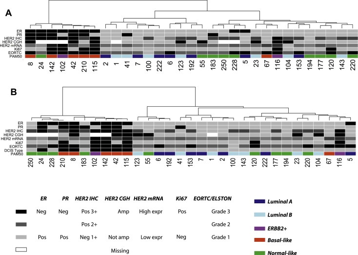
Ductal carcinoma in situ (DCIS) is a non-invasive form of breast cancer where cells restricted to the ducts exhibit an atypical phenotype. Some DCIS lesions are believed to rapidly transit to invasive ductal carcinomas (IDCs), while others remain unchanged. Existing classification systems for DCIS fail to identify those lesions that transit to IDC. We studied gene expression patterns of 31 pure DCIS, 36 pure invasive cancers and 42 cases of mixed diagnosis (invasive cancer with an in situ component) using Agilent Whole Human Genome Oligo Microarrays 44k. Six normal breast tissue samples were also included as controls. qRT-PCR was used for validation. All DCIS and invasive samples could be classified into the "intrinsic" molecular subtypes defined for invasive breast cancer. Hierarchical clustering establishes that samples group by intrinsic subtype, and not by diagnosis. We observed heterogeneity in the transcriptomes among DCIS of high histological grade and identified a distinct subgroup containing seven of the 31 DCIS samples with gene expression characteristics more similar to advanced tumours. A set of genes independent of grade, ER-status and HER2-status was identified by logistic regression that univariately classified a sample as belonging to this distinct DCIS subgroup. qRT-PCR of single markers clearly separated this DCIS subgroup from the other DCIS, and contains samples from several histopathological and intrinsic molecular subtypes. The genes that differentiate between these two types of DCIS suggest several processes related to the re-organisation of the microenvironment. This raises interesting possibilities for identification of DCIS lesions both with and without invasive characteristics, which potentially could be used in clinical assessment of a woman's risk of progression, and lead to improved management that would avoid the current over- and under-treatment of patients.
(c) 2010 Federation of European Biochemical Societies. Published by Elsevier B.V. All rights reserved.
Figures


References
-
- Allred, D.C. , Wu, Y. , Mao, S. , Nagtegaal, I.D. , Lee, S. , Perou, C.M. , Mohsin, S.K. , O'Connell, P. , Tsimelzon, A. , Medina, D. , 2008. Ductal carcinoma in situ and the emergence of diversity during breast cancer evolution. Clin. Cancer Res. 14, 370–378. - PubMed
-
- Blick, T. , Widodo, E. , Hugo, H. , Waltham, M. , Lenburg, M.E. , Neve, R.M. , Thompson, E.W. , 2008. Epithelial mesenchymal transition traits in human breast cancer cell lines. Clin. Exp. Metastasis. 25, 629–642. - PubMed
-
- Casey, T. , Bond, J. , Tighe, S. , Hunter, T. , Lintault, L. , Patel, O. , Eneman, J. , Crocker, A. , White, J. , Tessitore, J. , Stanley, M. , Harlow, S. , Weaver, D. , Muss, H. , Plaut, K. , 2009. Molecular signatures suggest a major role for stromal cells in development of invasive breast cancer. Breast Cancer Res. Treat. 114, 47–62. - PubMed
Publication types
MeSH terms
Substances
LinkOut - more resources
Full Text Sources
Other Literature Sources
Medical
Molecular Biology Databases
Research Materials
Miscellaneous
