. 2010 Sep;61(14):3935-45.
doi: 10.1093/jxb/erq210. Epub 2010 Jul 27.

Cloning, functional characterization, and co-expression studies of a novel aquaporin (FaPIP2;1) of strawberry fruit

Affiliations

Cloning, functional characterization, and co-expression studies of a novel aquaporin (FaPIP2;1) of strawberry fruit

Karina Alleva et al. J Exp Bot. 2010 Sep.

Abstract

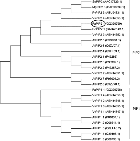
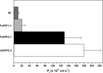
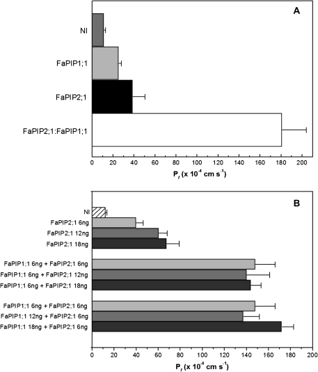
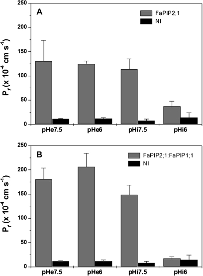
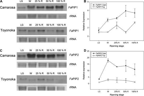
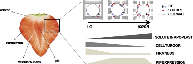
In strawberry, the putative participation of aquaporins should be considered during fruit ripening. Furthermore, the availability of different firmness cultivars in this non-climacteric fruit is a very useful tool to determine their involvement in softening. In a previous work, the cloning of a strawberry fruit-specific aquaporin, FaPIP1;1, which showed an expression profile associated with fruit ripening was reported. Here, FaPIP2;1, an aquaporin subtype of PIP2 was cloned and its functional characterization in Xenopus oocytes determined. The FaPIP2;1 gene encodes a water channel with high water permeability (P(f)) that is regulated by cytosolic pH. Interestingly, the co-expression of both FaPIP subtypes resulted in an enhancement of water permeability, showing P(f) values that exceeds their individual contribution. The expression pattern of both aquaporin subtypes in two cultivars with contrasting fruit firmness showed that the firmer cultivar (Camarosa) has a higher accumulation of FaPIP1 and FaPIP2 mRNAs during fruit ripening when compared with the softer cultivar (Toyonoka). In conclusion, not only FaPIP aquaporins showed an expression pattern associated with fruit firmness but it was also shown that the enhancement of water transfer through the plasma membrane is coupled to the presence/absence of the co-expression of both subtypes.


Figures

Fig. 1.
Fig. 1.
Phylogenetic analysis of full-length deduced amino acid sequences of plant aquaporins, including our clones, FaPIP2;1 and FaPIP1;1. Deduced amino acid sequences from full-length plant aquaporins encoding genes from Arabidopsis thaliana, Vitis vinifera, and other plant PIP2s with high identity to FaPIP2;1 were compared using Clustal W. Phylogenetic analyses were conducted using MEGA version 4 (Tamura et al., 2007).
Fig. 2.
Fig. 2.
Alignment of predicted amino acid sequence of Fragaria×ananassa aquaporin (FaPIP2;1) with other aquaporins (ClustalX). The predicted amino acid sequence of FaPIP2;1 was compared with aquaporins from different sources (aquaporins with the higher identity with FaPIP2;1 or very well studied in the literature). Transmembrane domains are shown with a dashed line below the alignment; triangles indicate a potential diacidic motif (putative ER signal); circles indicate the NPA selectivity filter; a star indicates His199, and inverted triangles putative phosphorylated Ser residues.
Fig. 3.
Fig. 3.
Functional expression of FaPIP2;1 in Xenopus oocytes. Calculated mean water permeabilities (Pf±SEM) of oocytes under hypo-osmotic conditions for NI (non-injected, negative control) or expressing 25 ng of FaPIP1;1, FaPIP2;1, and AtPIP2;3 (used as positive control) are shown. The number of measured oocytes for each condition is 5 to 8. Pf of oocytes expressing both FaPIP2;1 and AtPIP2;3 present significant differences from negative control (p <0.05).
Fig. 4.
Fig. 4.
Co-expression of FaPIP2;1 and FaPIP1;1. (A) Co-expression of 3 ng of cRNA of FaPIP2;1 with 12 ng of cRNA of FaPIP1;1 is shown (white bar). As a control, 3 ng of FaPIP2;1 (black bar) and 12 ng of FaPIP1;1 (light grey bar) were injected separately. The co-expression increased significantly the water permeability six times compared with the expression of FaPIP2;1 alone (p <0.05). Data are expressed as mean Pf±SEM, n=5 or 6 oocytes. NI are non-injected oocytes (grey bar). (B) Increasing cRNA mass of FaPIP2;1 injected alone in oocytes (from 6 ng to 18 ng) shows a increasing Pf. This relationship is not observed in FaPIP2;1-FaPIP1;1 co-expressing oocytes. Pf remains high but constant no matter the cRNA mass ratio injected. Data are shown as mean Pf ±SEM, n=6–8 oocytes.
Fig. 5.
Fig. 5.
Pf inhibitory response after cytosolic acidification. (A) FaPIP2;1 expressing oocytes were exposed to different external (pHe) or internal (pHi) pH conditions. Negative controls (NI) are non-injected oocytes. Pf for oocytes expressing FaPIP2;1 exposed to internal acidification is statistically different from its control (i.e. pHi=7.5), while treatment with pHe=6.0 does not result in a significant inhibition (p <0.05) when compared with its control (pHe=7.5). (B) FaPIP2;1-FaPIP1;1 was co-expressed in Xenopus oocytes and exposed to different external (pHe) or internal (pHi) pH conditions. Negative controls (NI) are non-injected oocytes. Data are shown as mean Pf±SEM, n=8–10 oocytes.
Fig. 6.
Fig. 6.
FaPIP1 and FaPIP2 expression pattern during strawberry fruit ripening. (A, C). Northern blot showing the accumulation of FaPIP mRNA in different ripening stages of strawberry fruits: large green (LG), white (W), 25% red (25%R), 50% red (50%R), and 100% red (100%R) for two different cultivars, a firm (Camarosa) and a softer one (Toyonoka). (B, D) Quantification of FaPIP1 and FaPIP2 expression relative to Toyonoka LG stage is shown for Camarosa and Toyonoka cultivars (Gel Pro Analizer v 3.0 was used). Data are shown as mean ±SEM, n=2.
Fig. 7.
Fig. 7.
Hypothetical and schematic representation of the role of aquaporins in fruit ripening. Section of a strawberry fruit showing fibrovascular strands (vascular bundles), achenes, the interior of the receptacle (pith), and main parenchyma; all marked tissues are possible targets of PIP expression due to their participation in water and solute balance in strawberry fruit.

References

    1. Agre P, Mathai JC, Smith BL, Preston GM. Functional analyses of aquaporin water channel proteins. Methods in Enzymology. 1999;294:550–572. - PubMed
    1. Aharoni A, Keizer LCP, Van Den Broeck HC, Blanco-Portales R, Muñoz-Blanco J, Bois G, Smit P, De Vos RCH, O'Connell AP. Novel insight into vascular, stress, and auxin-dependent and -independent gene expression programs in strawberry, a nonclimacteric fruit. Plant Physiology. 2002;129:1019–1031. - PMC - PubMed
    1. Altschul SF, Madden TL, Schaffer AA, Zhang J, Zhang Z, Miller W, Lipman DJ. Gapped BLAST and PSI-BLAST: a new generation of protein database search programs. Nucleic Acids Research. 1997;25:3389–3402. - PMC - PubMed
    1. Azad AK, Katsuhara M, Sawa Y, Ishikawa T, Shibata H. Characterization of four plasma membrane aquaporins in tulip petals: a putative homolog is regulated by phosphorylation. Plant and Cell Physiology. 2008;49:1196–1208. - PubMed
    1. Beitz E, Pavlovic-Djuranovic S, Yasui M, Agre P, Schultz JE. Molecular dissection of water and glycerol permeability of the aquaglyceroporin from Plasmodium falciparum by mutational analysis. Proceedings of the National Academy of Sciences, USA. 2004;101:1153–1158. - PMC - PubMed

Publication types