. 2010 Oct 1;285(40):30851-60.
doi: 10.1074/jbc.M110.110957. Epub 2010 Jul 27.

Phosphorylation of tau at Thr212, Thr231, and Ser262 combined causes neurodegeneration

Affiliations

Phosphorylation of tau at Thr212, Thr231, and Ser262 combined causes neurodegeneration

Alejandra D Alonso et al. J Biol Chem. .

Abstract

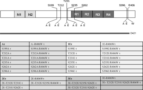
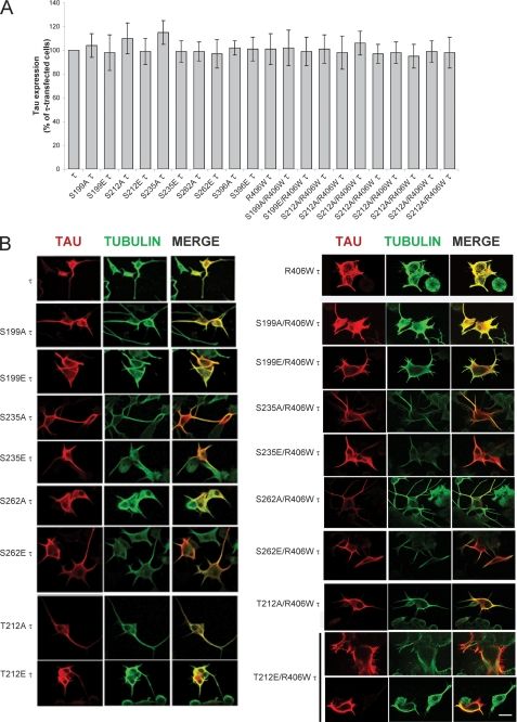
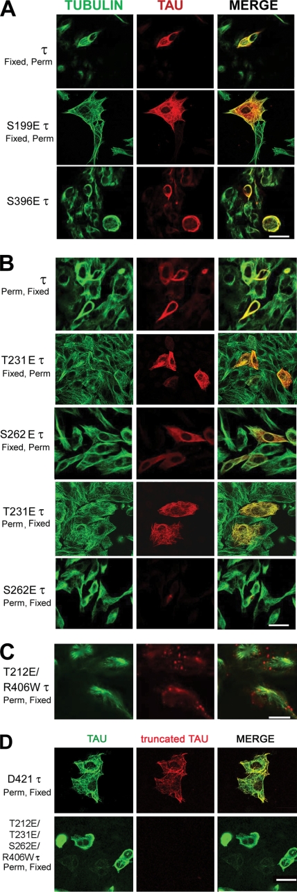
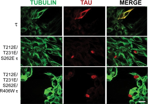
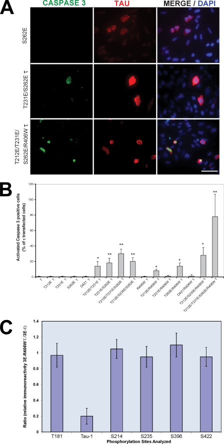
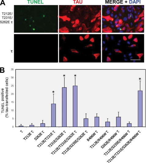
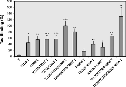
Abnormal hyperphosphorylation of the microtubule-associated protein Tau is a hallmark of Alzheimer disease and related diseases called tauopathies. As yet, the exact mechanism by which this pathology causes neurodegeneration is not understood. The present study provides direct evidence that Tau abnormal hyperphosphorylation causes its aggregation, breakdown of the microtubule network, and cell death and identifies phosphorylation sites involved in neurotoxicity. We generated pseudophosphorylated Tau proteins by mutating Ser/Thr to Glu and, as controls, to Ala. These mutations involved one, two, or three pathological phosphorylation sites by site-directed mutagenesis using as backbones the wild type or FTDP-17 mutant R406W Tau. Pseudophosphorylated and corresponding control Tau proteins were expressed transiently in PC12 and CHO cells. We found that a single phosphorylation site alone had little influence on the biological activity of Tau, except Thr(212), which, upon mutation to Glu in the R406W background, induced Tau aggregation in cells, suggesting phosphorylation at this site along with a modification on the C-terminal of the protein facilitates self-assembly of Tau. The expression of R406W Tau pseudophosphorylated at Thr(212), Thr(231), and Ser(262) triggered caspase-3 activation in as much as 85% of the transfected cells, whereas the corresponding value for wild type pseudophosphorylated Tau was 30%. Cells transfected with pseudophosphorylated Tau became TUNEL-positive.


Figures

FIGURE 1.
FIGURE 1.
Tau constructs employed for transfection of cells. N1 and N2 represent the two amino-terminal inserts, and R1, R2, R3, and R4 represent the four microtubule-binding domain repeats. Indicated are the S/T sites, which were pseudophosphorylated in wild type or R406W mutated Tau (τ) to Glu (E) and as control to Ala (A) in the present study. Asp421 Tau is shown (D421).
FIGURE 2.
FIGURE 2.
Expression of single-site pseudophosphorylated Tau (1E Tau and 1E R406W Tau) in PC12 cells. A, the levels of Tau (τ) expression in the cells was measured by quantifying the immunofluorescence of Tau relative to that of tubulin, using ImageJ software. The ratio of Tau to tubulin was considered 100% for cells transfected with wild type Tau. Error bars, SD. B, PC12 cells were transfected with pseudophosphorylated Tau for 24–48 h, and its expression was studied by immunocytochemistry as described. The cells were double labeled with 134d (Tau, red fluorescence) and DM1A (tubulin, green fluorescence). Merge is shown in yellow. Bar, 25 μm.
FIGURE 3.
FIGURE 3.
Expression of single-site pseudophosphorylated Tau (1E Tau and 1E R406W Tau) and Asp421 truncated Tau (Asp421 Tau and Asp421/R406W Tau) in CHO cells. A, cells were transfected with pseudophosphorylated or truncated Tau, and its expression was studied by immunocytochemistry at 24–48 h as described. The cells were double labeled with 134d (Tau, red fluorescence) and DM1A (tubulin, green fluorescence). Merge is shown in yellow. B, cells transfected with S262E Tau. The cells were fixed before (Fixed) or after (Perm) permeabilization. S262E Tau bound weakly to microtubules. C, cells transfected with T212E/R406W Tau. After 48 h, the cells were permeabilized with 0.1% Nonidet P-40 before fixed and processed for immunocytochemistry and double labeled with 134d (Tau, red) and DM1A (tubulin, green). T212E/R406W Tau appeared to form aggregates devoid of tubulin, decorating the microtubules. Bar, 25 μm. D, cells transfected with Asp421 Tau and double labeled with 134d (Tau, green fluorescence) and Tau C3 (truncated Tau, red fluorescence). Bar, 16 μm.
FIGURE 4.
FIGURE 4.
CHO cells transfected with either Tau or T212E/T231E/S262E Tau or T212E/T231E/S262E/R406W Tau. After 48 h, the cells were permeabilized with 0.1% Nonidet P-40 before fixing and then processed for immunocytochemistry and double labeled with 134d (Tau) and DM1A (tubulin). Merge is shown in yellow. 3E Tau appeared to localize in the nucleus, and tubulin staining was markedly reduced in the transfected cells. Bar, 25 μm.
FIGURE 5.
FIGURE 5.
Caspase activation in Tau-transfected CHO cells. A, cells were transfected with pseudophosphorylated wild type and R406W-mutated Tau proteins. After 48 h, the cells were double labeled with 134d (Tau) and activated caspase-3. Cells were counterstained with DAPI to visualize the nuclei. Bar, 50 μm. B, quantification of cells double stained for Tau and activated caspase-3. Approximately 300 cells were counted in each transfection. T212E/T231E/S262E/R406W Tau induced the highest caspase activation in the transfected cells. Error bars, SD. *, p < 0.05; **, p < 0.01; Error bars, SD. *, p < 0.05; **, p < 0.01. C, cells were transfected with T212E/T231E/S262E Tau or T212E/T231E/S262E/R406W Tau. (After 48 h, the cells were harvested, and phosphorylation of Tau at different sites (Thr181, Ser199/202 (Tau-1), Ser214, Ser235, Ser396, and Ser422) was determined by quantitative immuno-dot blots using antibodies specific to total and different phospho-Tau proteins.) The results are shown as the ratio of the immunoreactivity for T212E/T231E/S262E/R406W Tau (3E R406W Tau) to T212E/T231E/S262E Tau (3E Tau). No significant differences (p < 0.5) were found in the sites investigated between 3E Tau and 3E R406W Tau, with the exception of the Tau-1 site (reactive when Ser198/199–202 are not phosphorylated); Error bars, SD.
FIGURE 6.
FIGURE 6.
TUNEL staining in Tau-transfected CHO cells. A, CHO cells were transiently transfected with Tau and R406W Tau, and these Tau proteins pseudophosphorylated at various sites. After 48 h, the cells were double labeled with 134d (Tau) and TUNEL. Cells were counterstained with DAPI to visualize the nuclei. B, quantitation of cells double stained for Tau and TUNEL. The Tau construct used in each case is indicated. Approximately 300 cells were counted per Tau construct. T212E/T231E Tau, T231E/S262E Tau, T212E/T231E/S262E Tau, and T212E/T231E/S262E/R406W Tau induced apoptosis in the transfected cells. Bar, 50 μm. Error bars, SD. *, p < 0.05.
FIGURE 7.
FIGURE 7.
Pseudophosphorylated Tau binds normal Tau. CHO cells were transfected with different pseudophosphorylated Tau constructs, and cell extracts enriched in Tau were obtained as described under “Experimental Procedures.” Binding of normal Tau to pseudophosphorylated Tau was quantified as described under “Experimental Procedures.” The binding of Tau to T212E/T231E/S262E Tau was taken as 100%. Pseudophosphorylated Tau bound to normal Tau.

Similar articles

Cited by

References

    1. Grundke-Iqbal I., Iqbal K., Tung Y. C., Quinlan M., Wisniewski H. M., Binder L. I. (1986) Proc. Natl. Acad. Sci. U.S.A. 83, 4913–4917 - PMC - PubMed
    1. Weingarten M. D., Lockwood A. H., Hwo S. Y., Kirschner M. W. (1975) Proc. Natl. Acad. Sci. U.S.A. 72, 1858–1862 - PMC - PubMed
    1. Goedert M., Spillantini M. G., Jakes R., Rutherford D., Crowther R. A. (1989) Neuron 3, 519–526 - PubMed
    1. Himmler A., Drechsel D., Kirschner M. W., Martin D. W., Jr. (1989) Mol. Cell. Biol. 9, 1381–1388 - PMC - PubMed
    1. Goedert M., Spillantini M. G., Cairns N. J., Crowther R. A. (1992) Neuron 8, 159–168 - PubMed

Publication types