. 2010 Aug 15;123(Pt 16):2823-33.
doi: 10.1242/jcs.065565. Epub 2010 Jul 27.

SUMOylation modulates the function of Aurora-B kinase

Affiliations

SUMOylation modulates the function of Aurora-B kinase

Gonzalo Fernández-Miranda et al. J Cell Sci. .

Abstract

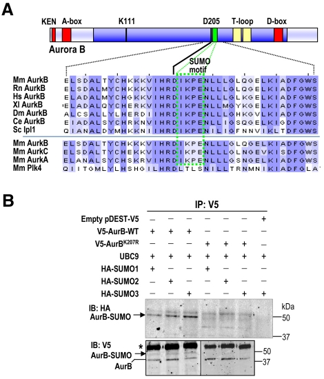
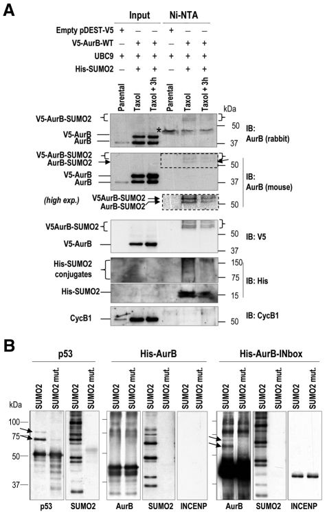
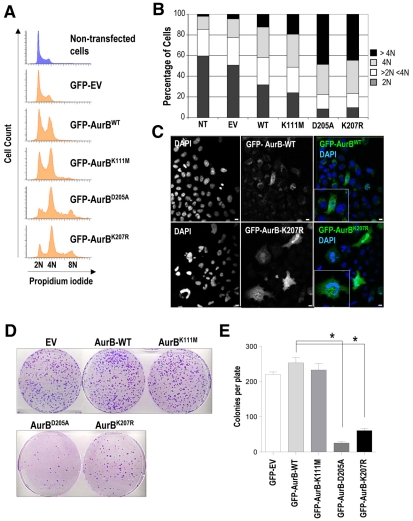
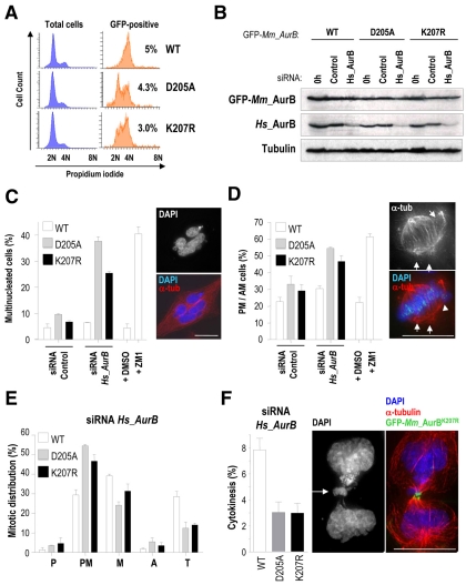
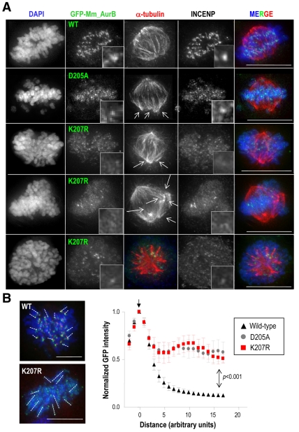
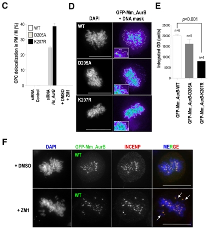
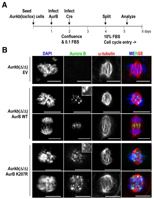
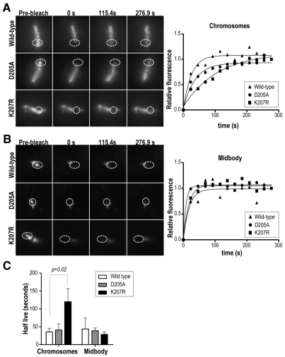
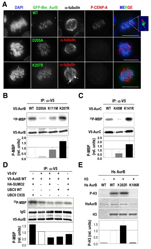
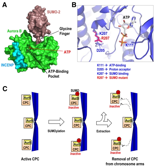
Aurora kinases are central regulators of mitotic-spindle assembly, chromosome segregation and cytokinesis. Aurora B is a member of the chromosomal passenger complex (CPC) with crucial functions in regulation of the attachment of kinetochores to microtubules and in cytokinesis. We report here that Aurora B contains a conserved SUMO modification motif within its kinase domain. Aurora B can bind SUMO peptides in vitro when bound to the IN-box domain of its CPC partner INCENP. Mutation of Lys207 to arginine (Aurora B(K207R)) impairs the formation of conjugates of Aurora B and SUMO in vivo. Expression of the SUMO-null form of Aurora B results in abnormal chromosome segregation and cytokinesis failure and it is not able to rescue mitotic defects in Aurora-B-knockout cells. These defects are accompanied by increased levels of the CPC on chromosome arms and defective centromeric function, as detected by decreased phosphorylation of the Aurora-B substrate CENP-A. The Aurora-B(K207R) mutant does not display reduced kinase activity, suggesting that functional defects are probably a consequence of the altered localization, rather than decreased intrinsic kinase activity. These data suggest that SUMOylation of Aurora B modulates its function, possibly by mediating the extraction of CPC complexes from chromosome arms during prometaphase.


Figures

Fig. 1.
Fig. 1.
A SUMOylation motif in Aurora B. (A) Multiple sequence alignment of Aurora B proteins from different species using Clustal software. Mm, Mus musculus; Rn, Rattus norvegicus; Hs, Homo sapiens; Xl, Xenopus laevis; Dm, Drosophila melanogaster; Ce, Caenorhabditis elegans; Sc, single Aurora family member (Ipl1) from Saccharomyces cerevisiae. The SUMO motif (green box) is highly conserved in all these Aurora-B proteins, as well as in Aurora A and Aurora C, but it is not present in Polo-like kinase 4 (Plk4), a close mitotic relative of these mitotic kinases. The kinase domain is shown in dark blue, whereas the degradation motifs (KEN, A- and D-boxes) are represented as red boxes. Note that the SUMO motif is located one residue downstream of the aspartate (D205) that is crucial for ATP binding. Residue numbers correspond to the mouse Aurora-B protein. (B) In vivo SUMOylation of Aurora B at K207. HEK293 asynchronous cells were transiently transfected with vectors expressing V5-Aurora-B WT, Aurora BK207R, HA-SUMO1, -SUMO2 or -SUMO3 and the E2 SUMO ligase UBC9, as indicated. The empty vector pDEST-V5 was used as a negative control. V5-Aurora-B proteins were immunoprecipitated (IP) 48 hours after transfection using anti-V5 antibodies and immunoblotted (IB) with antibodies against the HA and V5 tags. Aurora-B-SUMO conjugates are found after detection of the HA and V5 tags in cells transfected with WT V5-Aurora-B but not in cells transfected with the V5-Aurora-BK207R mutant form. V5-Aurora-B immunoprecipitated in all lanes. Mouse IgGs are indicated with an asterisk.
Fig. 2.
Fig. 2.
In vivo and in vitro SUMOylation of Aurora B. (A) HeLa-SUMO2 or parental cells were transfected with UBC9 or V5-Aurora-B-expressing vectors as indicated. Cells were harvested after 18 hours in the presence of taxol or 3 hours after release from taxol. His-SUMO2 conjugates were recovered on Ni-NTA beads and analyzed by immunoblotting (IB). The indicated antibodies were used for immunoblot analysis on total protein lysates (input) or SUMO2 conjugates purified with Ni-NTA beads. The position of V5-Aurora-B-SUMO2 conjugates (~60 kDa and higher) is indicated by brackets. An additional band of ~55 kDa (predicted size for SUMO conjugates with endogenous Aurora B) is detected by the mouse monoclonal antibody against Aurora B. The asterisk indicates an unspecific band detected by the rabbit, but not the mouse, anti-Aurora-B antibody. (B) In vitro SUMOylation of Aurora B. Recombinant p53, Aurora B or Aurora B in complex with the IN-box of INCENP were incubated in the presence of SUMOylation machinery and SUMO2 or a SUMO2 non-conjugatable mutant. Proteins were immunodetected using specific antibodies against the indicated proteins. p53- or Aurora-B-SUMO2 conjugates are indicated by arrows.
Fig. 3.
Fig. 3.
Aurora BK207R induces polyploidy and defective cell viability. (A) DNA content of HEK293 cells transiently transfected with a GFP empty vector (EV) and GFP-tagged vectors for wild-type Aurora B and Aurora B mutants (Aurora BK111M, Aurora BD205A and Aurora BK207R) and fixed 72 hours after transfection. HEK293 GFP-positive cells were detected and used for cell-cycle analysis. Overexpression of a SUMO-null Aurora-B protein (Aurora BK207R) deregulates cell-cycle progression inducing polyploidy in the same way as Aurora-BD205A kinase-dead form. (B) Bar graph quantification of cellular DNA content, showing a dramatic increase of polyploidy in cells overexpressing Aurora BD205A and Aurora BK207R mutants. (C) Immunofluorescence (GFP, green; DAPI, blue) images revealing that cells overexpressing Aurora BK207R are mostly multinucleated and display nuclear aberrancies. Scale bars: 10 μm. (D) Colony-formation assay in U2OS cells stably expressing GFP empty vector (EV) and GFP-tagged vectors for Aurora B (wild-type and mutants). U2OS GFP-positive cells were sorted 48 hours after transfection, replated in the presence of 1 mg/ml G418, then fixed 12 days later and stained with Crystal Violet to determine colony number. 50,000 GFP-positive cells were sorted and plated in 10 cm plates. (E) Quantification of the number of macroscopic colonies per 10 cm plate showing a significant (*P<0.001) reduction in the number of colonies in cells expressing Aurora BD205A and Aurora BK207R.
Fig. 4.
Fig. 4.
Defective mitotic progression and cytokinesis in the presence of Aurora BK207R. HeLa cells stably expressing siRNA-resistant mouse Aurora B fused to GFP were nucleofected with human AURKB siRNA oligonucleotides or a control siRNA and analyzed 36 hours after nucleofection by immunoblotting and immunofluorescence. (A) Generation and validation of HeLa stable cell lines by cytometry cell-cycle analysis. The analysis of the total cell population is shown in blue histograms, whereas the analysis of the GFP-positive population is shown in orange histograms accompanied by the percentage of these cells over the whole population. At least three different stable clones were analyzed for each GFP-Aurora-B fusion protein. Depicted histograms are from representative stable clones. (B) Western blot analysis showing efficient depletion of human endogenous Aurora B and protein levels of siRNA-resistant mouse GFP-Aurora-B proteins in HeLa stable cell lines. Tubulin was used as loading control. (C) Quantification of the percentage of interphase multinucleated cells. Stable expression of GFP-Aurora-BD205A and Aurora-BK207R induces accumulation of multinucleated cells (see picture) upon depletion of endogenous Aurora B. Around 1000 interphase cells were counted per point. (D) Quantification of the percentage of mitotic cells in prometaphase (PM) or abnormal metaphases (AM) with misaligned chromosomes (arrowheads) and multiple poles (arrows) revealing a significant arrest at these stages in cells lacking the endogenous Aurora B and stably expressing GFP-AuroraBD205A and -AuroraBK207R. About 50-100 mitotic cells were counted per point. Chemical inhibition of Aurora-B kinase activity with ZM447439 (ZM1) was included in C and D as a positive control. (E) Mitotic distribution of stable cell lines after depletion of endogenous Aurora B. Expression of GFP-Aurora-BD205A and -Aurora-BK207R provokes an arrest at prometaphase and a reduction in cells at telophase. P, prophase; PM, prometaphase; M, metaphase; A, anaphase; T, telophase. (F) GFP-Aurora-BD205A and -Aurora-BK207R stable cell lines display a reduced number of cells undergoing cytokinesis. In these mutant cells, the remaining cytokinesis figures are usually abnormal with chromosomal bridges (arrow). α-tubulin (α-tub) is in red, GFP-Aurora-B in green and DAPI (DNA) in blue. Scale bars: 10 μm.
Fig. 5.
Fig. 5.
The Aurora-B K207 residue is required for proper localization of CPC proteins at the centromere. (A) Immunofluorescence images of HeLa stable cell lines after depletion of endogenous Aurora B. Wild-type Aurora B colocalizes with INCENP at the centromeric regions. However, GFP-Aurora-BD205A and -Aurora-BK207R mutants display a disperse localization over the chromatin that also affects to INCENP in PM/M cells. Multipolar spindles are also present in these mutant cells (arrows). Scale bars: 10 μm. (B) Quantification of GFP-Aurora-B at chromosome arms from the centromere (Position 0; vertical arrow). Results are means ± s.e.m. Scale bars: 5 μm. (C) Quantification of the percentage of PM/M cells in which GFP-Aurora-B fusions and INCENP are delocalized. Delocalization of Aurora B is only observed in Aurora-BD205A and Aurora-BK207R mutants after depletion of the endogenous protein. At least 50 mitotic cells were counted per point. (D) Images pseudo-coloured with Metamorph software to visualize the distribution of GFP-Aurora-B signal in stable cell lines after depletion of endogenous Aurora B. Intensity of the signal is represented in a colour scale: (low) purple, blue, green, yellow, red (high). The contour of DNA (DAPI staining) is delineated. Scale bars: 10 μm. (E) Quantification of the integrated optical density (OD) of GFP-Aurora-B signal over the chromatin shows a significant reduction (P<0.0001) of GFP-Aurora-BK207R, but not -Aurora-BD205A, signal density. (F) Chemical inhibition of Aurora B with ZM1 does not delocalize the protein although it provokes chromosome misalignment (arrows). α-tubulin is in red in A, INCENP is in red in E, GFP-Aurora B is in green and DAPI (DNA) in blue. Scale bars: 10 μm.
Fig. 5.
Fig. 5.
The Aurora-B K207 residue is required for proper localization of CPC proteins at the centromere. (A) Immunofluorescence images of HeLa stable cell lines after depletion of endogenous Aurora B. Wild-type Aurora B colocalizes with INCENP at the centromeric regions. However, GFP-Aurora-BD205A and -Aurora-BK207R mutants display a disperse localization over the chromatin that also affects to INCENP in PM/M cells. Multipolar spindles are also present in these mutant cells (arrows). Scale bars: 10 μm. (B) Quantification of GFP-Aurora-B at chromosome arms from the centromere (Position 0; vertical arrow). Results are means ± s.e.m. Scale bars: 5 μm. (C) Quantification of the percentage of PM/M cells in which GFP-Aurora-B fusions and INCENP are delocalized. Delocalization of Aurora B is only observed in Aurora-BD205A and Aurora-BK207R mutants after depletion of the endogenous protein. At least 50 mitotic cells were counted per point. (D) Images pseudo-coloured with Metamorph software to visualize the distribution of GFP-Aurora-B signal in stable cell lines after depletion of endogenous Aurora B. Intensity of the signal is represented in a colour scale: (low) purple, blue, green, yellow, red (high). The contour of DNA (DAPI staining) is delineated. Scale bars: 10 μm. (E) Quantification of the integrated optical density (OD) of GFP-Aurora-B signal over the chromatin shows a significant reduction (P<0.0001) of GFP-Aurora-BK207R, but not -Aurora-BD205A, signal density. (F) Chemical inhibition of Aurora B with ZM1 does not delocalize the protein although it provokes chromosome misalignment (arrows). α-tubulin is in red in A, INCENP is in red in E, GFP-Aurora B is in green and DAPI (DNA) in blue. Scale bars: 10 μm.
Fig. 6.
Fig. 6.
Aurora BK207R does not rescue mitotic defects in Aurora-B-knockout MEFs. (A) Schematic representation of the protocol followed for rescue upon acute deletion of Aurora B in quiescent cells. Primary (passage 2) Aurkb(lox/lox) MEFs were seeded at 80% confluence and infected the following day with retroviruses expressing Aurora-BWT or Aurora-BK207R. Next day, cultures reach confluence and were cultured in 0.1% FBS and infected with adenoviruses expressing Cre recombinase (AdCre). Two days after the infection, cells were split in new plates at low confluence in the presence of 10% FBS to induce entry into the cell cycle. Mitotic cells were analyzed by immunofluorescence during the first round of division (28 hours after splitting). (B) Acute deletion of Aurora B in MEFs provokes chromosome misalignment and spindle defects that are rescued by exogenous, properly localized, wild-type Aurora B [Aurkb(Δ/Δ); Aurora BWT; green signal in merged images]. The exogenous Aurora BK207R, however, remains diffuse on chromosome arms and it does not rescue the Aurkb-null defects resulting in chromosome misalignment and impaired cytokinesis in Aurkb(Δ/Δ); Aurora BK207R cells. DNA (DAPI; blue) is delineated in the insets. α-tubulin is in red in merged images. Scale bars: 10 μm.
Fig. 7.
Fig. 7.
Defective dynamic localization of Aurora-B K207R at metaphase chromosomes. HeLa cells stably expressing GFP-tagged Aurora-B isoforms were used in a series of fluorescence recovery after photobleaching (FRAP) experiments. (A) Representative cases of recovery of wild-type Aurora B, Aurora BK207R and Aurora BD205A at the metaphase chromosomes. Delocalization of the Aurora-B mutant isoforms is not obvious as in Fig. 4 because these cells express the endogenous Aurora-B protein. (B) Representative images and quantification of similar FRAP assays at the mid-body. Graphs in both cases show the curves obtained using a regression analysis to integrate data from each stable clone. (C) The half-lives of GFP-Aurora-B at metaphase chromosomes and mid-bodies were compared using a t-test showing significant differences between the AurBK207R and the wild-type forms. Results are means ± s.e.m.
Fig. 8.
Fig. 8.
Centromeric mislocalization of Aurora-BK207R mutant prevents CENP-A phosphorylation without reducing its kinase activity. (A) Immunofluorescence images from HeLa stable cell lines treated with siRNA against endogenous Aurora B. GFP-Aurora-BD205A and -Aurora-BK207R mutants accumulate along chromosome arms and fail to phosphorylate CENP-A. α-tubulin staining is in red (only merged in the mutant forms), phosphorylated CENP-A, if present, is in red (only merged in the wild-type forms), GFP-Aurora-B in green and DAPI (DNA) in blue. The inset represents proper phosphorylation of CENP-A (red) by centromeric Aurora B (green). Scale bars: 10 μm. (B) In vitro kinase activity toward recombinant MBP of V5-tagged Aurora B proteins. HEK293 cells were transiently transfected with the indicated V5-Aurora-B expression vectors and the resulting exogenous expressed proteins were immunoprecipitated with anti-V5 antibody. The kinase activity of a SUMO-null (Aurora BK207R) Aurora B form is not reduced but partially increased. Aurora-B kinase-dead mutants K111M and D205A were included as negative controls of MBP phosphorylation. Quantification of [32P]MBP signal was calculated considering the total amount of V5-Aurora-B protein in each case. (C) Similar kinase assays using Aurora C constructs with the corresponding kinase-dead (Aurora CK45M) or SUMO-null (Aurora CK141R) mutations. (D) In vitro kinase assay in the presence of SUMO2, UBC9 or a dominant-negative UBC9 form (C93S mutant). Whereas the overexpression of SUMO2 and UBC9 results in decreased activity, the presence of UBC9 C93S results in increased Aurora-B-kinase activity in this assay. Data were normalized to the intensity of phosphorylated MBP in cells transfected only with wild-type Aurora B. (E) Phosphorylation of histone H3 (P-H3) by recombinant human wild-type (WT) Aurora B or the kinase-dead (K106R) and SUMO-null (K202R) mutants. Data were normalized to total levels of human Aurora B (HsAurB).
Fig. 9.
Fig. 9.
Modelling the Aurora-B-SUMO covalent interaction. (A) The crystal structures of human SUMO2 (Reverter and Lima, 2006) (PDB 2I03) and Xenopus Aurora B in complex with IN-box segment of INCENP (Sessa et al., 2005) (PDB 2BFY) were used to simulate the interaction between these molecules. SUMO2 might covalently bind the Aurora-B K207 residue (K218 in Xenopus crystal structure) through a finger composed of two glycine residues. SUMOylation is not predicted to affect binding of the Aurora-B partner and activator INCENP, but it might disturb the transfer of phosphate groups to its substrates. (B) Details of the ATP-binding pocket showing the localization of K111, D205 and K207 residues (K122, D216 and K218 in Xenopus, respectively) used in this study. The position of the ATP molecule was modelled after a superposition with the Aurora-A-TPX2 structure (PDB 1OL5). (C) A model for the role of SUMOylation of Aurora B in CPC function. SUMOylation of Aurora B might result in decreased kinase activity, probably as a consequence of interference of the SUMO residue with kinase function. In addition, the presence of the SUMO residue might trigger an unknown pathway that favours the removal of Aurora B and the CPC from chromatin. MT, microtubule.

References

    1. Adams R. R., Wheatley S. P., Gouldsworthy A. M., Kandels-Lewis S. E., Carmena M., Smythe C., Gerloff D. L., Earnshaw W. C. (2000). INCENP binds the Aurora-related kinase AIRK2 and is required to target it to chromosomes, the central spindle and cleavage furrow. Curr. Biol. 10, 1075-1078 - PubMed
    1. Ayaydin F., Dasso M. (2004). Distinct in vivo dynamics of vertebrate SUMO paralogues. Mol. Biol. Cell 15, 5208-5218 - PMC - PubMed
    1. Azuma Y., Dasso M. (2002). A new clue at the nuclear pore: RanBP2 is an E3 enzyme for SUMO1. Dev. Cell 2, 130-131 - PubMed
    1. Azuma Y., Arnaoutov A., Anan T., Dasso M. (2005). PIASy mediates SUMO-2 conjugation of Topoisomerase-II on mitotic chromosomes. EMBO J. 24, 2172-2182 - PMC - PubMed
    1. Bishop J. D., Schumacher J. M. (2002). Phosphorylation of the carboxyl terminus of inner centromere protein (INCENP) by the Aurora B Kinase stimulates Aurora B kinase activity. J. Biol. Chem. 277, 27577-27580 - PMC - PubMed

Publication types

MeSH terms