. 2010 Sep;256(3):827-35.
doi: 10.1148/radiol.10091890. Epub 2010 Jul 27.

Effect of computer-aided detection for CT colonography in a multireader, multicase trial

Affiliations

Effect of computer-aided detection for CT colonography in a multireader, multicase trial

Abraham H Dachman et al. Radiology. 2010 Sep.

Abstract

Purpose: To assess the effect of using computer-aided detection (CAD) in second-read mode on readers' accuracy in interpreting computed tomographic (CT) colonographic images.

Materials and methods: The contributing institutions performed the examinations under approval of their local institutional review board, with waiver of informed consent, for this HIPAA-compliant study. A cohort of 100 colonoscopy-proved cases was used: In 52 patients with findings positive for polyps, 74 polyps of 6 mm or larger were observed in 65 colonic segments; in 48 patients with findings negative for polyps, no polyps were found. Nineteen blinded readers interpreted each case at two different times, with and without the assistance of a commercial CAD system. The effect of CAD was assessed in segment-level and patient-level receiver operating characteristic (ROC) curve analyses.

Results: Thirteen (68%) of 19 readers demonstrated higher accuracy with CAD, as measured with the segment-level area under the ROC curve (AUC). The readers' average segment-level AUC with CAD (0.758) was significantly greater (P = .015) than the average AUC in the unassisted read (0.737). Readers' per-segment, per-patient, and per-polyp sensitivity for all polyps of 6 mm or larger was higher (P < .011, .007, .005, respectively) for readings with CAD compared with unassisted readings (0.517 versus 0.465, 0.521 versus 0.466, and 0.477 versus 0.422, respectively). Sensitivity for patients with at least one large polyp of 10 mm or larger was also higher (P < .047) with CAD than without (0.777 versus 0.743). Average reader sensitivity also improved with CAD by more than 0.08 for small adenomas. Use of CAD reduced specificity of readers by 0.025 (P = .05).

Conclusion: Use of CAD resulted in a significant improvement in overall reader performance. CAD improves reader sensitivity when measured per segment, per patient, and per polyp for small polyps and adenomas and also reduces specificity by a small amount.


Conflict of interest statement

See Materials and Methods for pertinent disclosures.

Figures

Figure 1a:
Figure 1a:
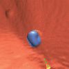
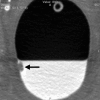
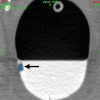
A 12-mm polyp (arrow) located in the rectum in 71-year-old woman and found with CAD and by nearly all readers at unassisted and CAD-assisted reads. (a) Prone axial image with CAD off shows polyp as a soft-tissue lesion on right rectal wall coated with oral contrast material. (b) Same axial 2D image with CAD on shows mark (blue), painting with a yellow circle. (c) Corresponding 3D image with CAD on shows polyp painted blue. (d) Supine axial 2D image shows polyp submerged in well-tagged residual fluid with CAD off and (e) same image with CAD on. Note that residual tagged fluid does not prevent CAD from detecting the polyp.
Figure 1b:
Figure 1b:
A 12-mm polyp (arrow) located in the rectum in 71-year-old woman and found with CAD and by nearly all readers at unassisted and CAD-assisted reads. (a) Prone axial image with CAD off shows polyp as a soft-tissue lesion on right rectal wall coated with oral contrast material. (b) Same axial 2D image with CAD on shows mark (blue), painting with a yellow circle. (c) Corresponding 3D image with CAD on shows polyp painted blue. (d) Supine axial 2D image shows polyp submerged in well-tagged residual fluid with CAD off and (e) same image with CAD on. Note that residual tagged fluid does not prevent CAD from detecting the polyp.
Figure 1c:
Figure 1c:
A 12-mm polyp (arrow) located in the rectum in 71-year-old woman and found with CAD and by nearly all readers at unassisted and CAD-assisted reads. (a) Prone axial image with CAD off shows polyp as a soft-tissue lesion on right rectal wall coated with oral contrast material. (b) Same axial 2D image with CAD on shows mark (blue), painting with a yellow circle. (c) Corresponding 3D image with CAD on shows polyp painted blue. (d) Supine axial 2D image shows polyp submerged in well-tagged residual fluid with CAD off and (e) same image with CAD on. Note that residual tagged fluid does not prevent CAD from detecting the polyp.
Figure 1d:
Figure 1d:
A 12-mm polyp (arrow) located in the rectum in 71-year-old woman and found with CAD and by nearly all readers at unassisted and CAD-assisted reads. (a) Prone axial image with CAD off shows polyp as a soft-tissue lesion on right rectal wall coated with oral contrast material. (b) Same axial 2D image with CAD on shows mark (blue), painting with a yellow circle. (c) Corresponding 3D image with CAD on shows polyp painted blue. (d) Supine axial 2D image shows polyp submerged in well-tagged residual fluid with CAD off and (e) same image with CAD on. Note that residual tagged fluid does not prevent CAD from detecting the polyp.
Figure 1e:
Figure 1e:
A 12-mm polyp (arrow) located in the rectum in 71-year-old woman and found with CAD and by nearly all readers at unassisted and CAD-assisted reads. (a) Prone axial image with CAD off shows polyp as a soft-tissue lesion on right rectal wall coated with oral contrast material. (b) Same axial 2D image with CAD on shows mark (blue), painting with a yellow circle. (c) Corresponding 3D image with CAD on shows polyp painted blue. (d) Supine axial 2D image shows polyp submerged in well-tagged residual fluid with CAD off and (e) same image with CAD on. Note that residual tagged fluid does not prevent CAD from detecting the polyp.
Figure 2a:
Figure 2a:
An 8-mm polyp (arrow) located in the ascending colon was visible only on supine 3D fly-through image (at 120° viewing angle) and only in antegrade direction in 65-year-old woman. The polyp was missed by all readers and with CAD. (a–d) Contiguous axial 2D images show polyp abutting a fold and partially submerged in tagged fluid; on the cut above (not shown), polyp abutted a normal fold. (e) Corresponding supine 3D retrograde endoluminal view shows the polyp located on the proximal side of a fold, explaining why it could not be seen on the retrograde view (ie, rectum to cecum, fly-through). Green line = computer-generated center line. (f, g) Prone axial 2D images in CT colonographic settings of (f) window width of 2000 HU and window level of 0 HU and (g) window width of 600 HU and window level of 0 HU show polyp with some tagging on the surface, making it easy to confuse for stool. (h) Prone view with the perspective optimized manually to show the polyp (arrows).
Figure 2b:
Figure 2b:
An 8-mm polyp (arrow) located in the ascending colon was visible only on supine 3D fly-through image (at 120° viewing angle) and only in antegrade direction in 65-year-old woman. The polyp was missed by all readers and with CAD. (a–d) Contiguous axial 2D images show polyp abutting a fold and partially submerged in tagged fluid; on the cut above (not shown), polyp abutted a normal fold. (e) Corresponding supine 3D retrograde endoluminal view shows the polyp located on the proximal side of a fold, explaining why it could not be seen on the retrograde view (ie, rectum to cecum, fly-through). Green line = computer-generated center line. (f, g) Prone axial 2D images in CT colonographic settings of (f) window width of 2000 HU and window level of 0 HU and (g) window width of 600 HU and window level of 0 HU show polyp with some tagging on the surface, making it easy to confuse for stool. (h) Prone view with the perspective optimized manually to show the polyp (arrows).
Figure 2c:
Figure 2c:
An 8-mm polyp (arrow) located in the ascending colon was visible only on supine 3D fly-through image (at 120° viewing angle) and only in antegrade direction in 65-year-old woman. The polyp was missed by all readers and with CAD. (a–d) Contiguous axial 2D images show polyp abutting a fold and partially submerged in tagged fluid; on the cut above (not shown), polyp abutted a normal fold. (e) Corresponding supine 3D retrograde endoluminal view shows the polyp located on the proximal side of a fold, explaining why it could not be seen on the retrograde view (ie, rectum to cecum, fly-through). Green line = computer-generated center line. (f, g) Prone axial 2D images in CT colonographic settings of (f) window width of 2000 HU and window level of 0 HU and (g) window width of 600 HU and window level of 0 HU show polyp with some tagging on the surface, making it easy to confuse for stool. (h) Prone view with the perspective optimized manually to show the polyp (arrows).
Figure 2d:
Figure 2d:
An 8-mm polyp (arrow) located in the ascending colon was visible only on supine 3D fly-through image (at 120° viewing angle) and only in antegrade direction in 65-year-old woman. The polyp was missed by all readers and with CAD. (a–d) Contiguous axial 2D images show polyp abutting a fold and partially submerged in tagged fluid; on the cut above (not shown), polyp abutted a normal fold. (e) Corresponding supine 3D retrograde endoluminal view shows the polyp located on the proximal side of a fold, explaining why it could not be seen on the retrograde view (ie, rectum to cecum, fly-through). Green line = computer-generated center line. (f, g) Prone axial 2D images in CT colonographic settings of (f) window width of 2000 HU and window level of 0 HU and (g) window width of 600 HU and window level of 0 HU show polyp with some tagging on the surface, making it easy to confuse for stool. (h) Prone view with the perspective optimized manually to show the polyp (arrows).
Figure 2e:
Figure 2e:
An 8-mm polyp (arrow) located in the ascending colon was visible only on supine 3D fly-through image (at 120° viewing angle) and only in antegrade direction in 65-year-old woman. The polyp was missed by all readers and with CAD. (a–d) Contiguous axial 2D images show polyp abutting a fold and partially submerged in tagged fluid; on the cut above (not shown), polyp abutted a normal fold. (e) Corresponding supine 3D retrograde endoluminal view shows the polyp located on the proximal side of a fold, explaining why it could not be seen on the retrograde view (ie, rectum to cecum, fly-through). Green line = computer-generated center line. (f, g) Prone axial 2D images in CT colonographic settings of (f) window width of 2000 HU and window level of 0 HU and (g) window width of 600 HU and window level of 0 HU show polyp with some tagging on the surface, making it easy to confuse for stool. (h) Prone view with the perspective optimized manually to show the polyp (arrows).
Figure 2f:
Figure 2f:
An 8-mm polyp (arrow) located in the ascending colon was visible only on supine 3D fly-through image (at 120° viewing angle) and only in antegrade direction in 65-year-old woman. The polyp was missed by all readers and with CAD. (a–d) Contiguous axial 2D images show polyp abutting a fold and partially submerged in tagged fluid; on the cut above (not shown), polyp abutted a normal fold. (e) Corresponding supine 3D retrograde endoluminal view shows the polyp located on the proximal side of a fold, explaining why it could not be seen on the retrograde view (ie, rectum to cecum, fly-through). Green line = computer-generated center line. (f, g) Prone axial 2D images in CT colonographic settings of (f) window width of 2000 HU and window level of 0 HU and (g) window width of 600 HU and window level of 0 HU show polyp with some tagging on the surface, making it easy to confuse for stool. (h) Prone view with the perspective optimized manually to show the polyp (arrows).
Figure 2g:
Figure 2g:
An 8-mm polyp (arrow) located in the ascending colon was visible only on supine 3D fly-through image (at 120° viewing angle) and only in antegrade direction in 65-year-old woman. The polyp was missed by all readers and with CAD. (a–d) Contiguous axial 2D images show polyp abutting a fold and partially submerged in tagged fluid; on the cut above (not shown), polyp abutted a normal fold. (e) Corresponding supine 3D retrograde endoluminal view shows the polyp located on the proximal side of a fold, explaining why it could not be seen on the retrograde view (ie, rectum to cecum, fly-through). Green line = computer-generated center line. (f, g) Prone axial 2D images in CT colonographic settings of (f) window width of 2000 HU and window level of 0 HU and (g) window width of 600 HU and window level of 0 HU show polyp with some tagging on the surface, making it easy to confuse for stool. (h) Prone view with the perspective optimized manually to show the polyp (arrows).
Figure 2h:
Figure 2h:
An 8-mm polyp (arrow) located in the ascending colon was visible only on supine 3D fly-through image (at 120° viewing angle) and only in antegrade direction in 65-year-old woman. The polyp was missed by all readers and with CAD. (a–d) Contiguous axial 2D images show polyp abutting a fold and partially submerged in tagged fluid; on the cut above (not shown), polyp abutted a normal fold. (e) Corresponding supine 3D retrograde endoluminal view shows the polyp located on the proximal side of a fold, explaining why it could not be seen on the retrograde view (ie, rectum to cecum, fly-through). Green line = computer-generated center line. (f, g) Prone axial 2D images in CT colonographic settings of (f) window width of 2000 HU and window level of 0 HU and (g) window width of 600 HU and window level of 0 HU show polyp with some tagging on the surface, making it easy to confuse for stool. (h) Prone view with the perspective optimized manually to show the polyp (arrows).

References

    1. Soto JA, Barish MA, Yee J. Reader training in CT colonography: how much is enough? Radiology 2005;237(1):26–27 - PubMed
    1. Dachman AH, Kelly KB, Zintsmaster MP, et al. Formative evaluation of standardized training for CT colonographic image interpretation by novice readers. Radiology 2008;249(1):167–177 - PubMed
    1. Mulhall BP, Veerappan GR, Jackson JL. Meta-analysis: computed tomographic colonography. Ann Intern Med 2005;142(8):635–650 - PubMed
    1. Summers RM, Jerebko AK, Franaszek M, Malley JD, Johnson CD. Colonic polyps: complementary role of computer-aided detection in CT colonography. Radiology 2002;225(2):391–399 - PubMed
    1. Bogoni L, Cathier P, Dundar M, et al. Computer-aided detection (CAD) for CT colonography: a tool to address a growing need. Br J Radiol 2005;78(spec no 1):S57–S62 - PubMed