. 2010 Nov 11;116(19):3899-906.
doi: 10.1182/blood-2009-12-257378. Epub 2010 Jul 27.

An epigenetic chromatin remodeling role for NFATc1 in transcriptional regulation of growth and survival genes in diffuse large B-cell lymphomas

Affiliations

An epigenetic chromatin remodeling role for NFATc1 in transcriptional regulation of growth and survival genes in diffuse large B-cell lymphomas

Lan V Pham et al. Blood. .

Abstract

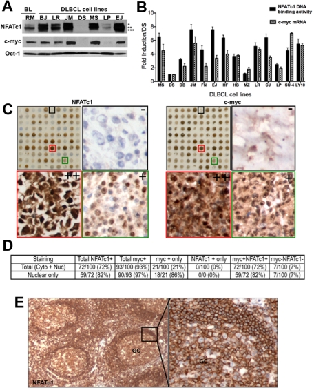
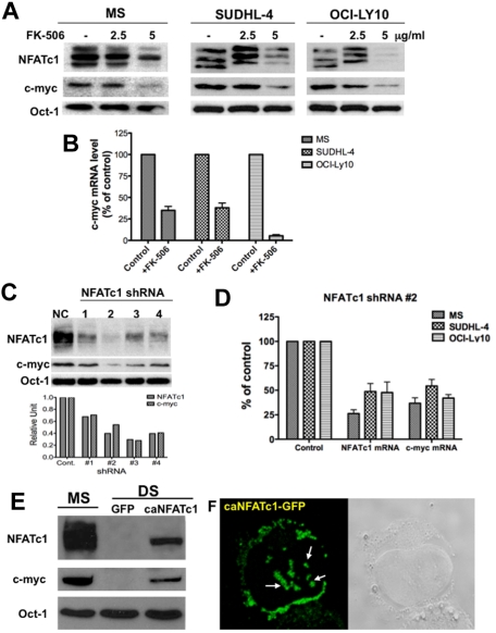
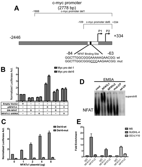
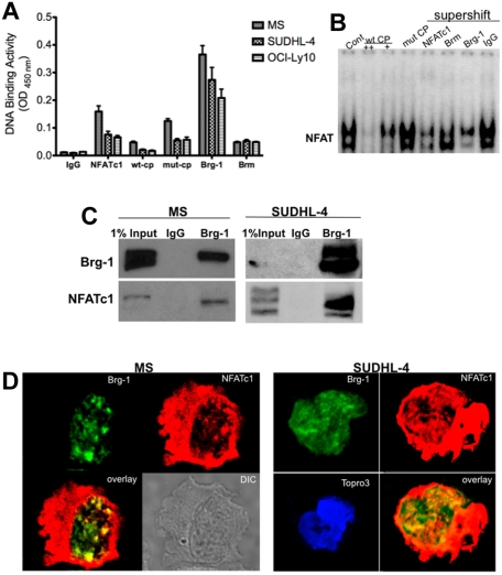
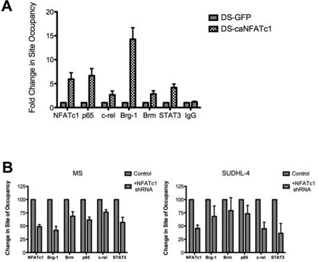
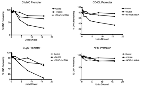
The nuclear factor of activated T cells (NFAT) family of transcription factors functions as integrators of multiple signaling pathways by binding to chromatin in combination with other transcription factors and coactivators to regulate genes central for cell growth and survival in hematopoietic cells. Recent experimental evidence has implicated the calcineurin/NFAT signaling pathway in the pathogenesis of various malignancies, including diffuse large B-cell lymphoma (DLBCL). However, the molecular mechanism(s) underlying NFATc1 regulation of genes controlling lymphoma cell growth and survival is still unclear. In this study, we demonstrate that the transcription factor NFATc1 regulates gene expression in DLBCL cells through a chromatin remodeling mechanism that involves recruitment of the SWItch/Sucrose NonFermentable chromatin remodeling complex ATPase enzyme SMARCA4 (also known as Brahma-related gene 1) to NFATc1 targeted gene promoters. The NFATc1/Brahma-related gene 1 complex induces promoter DNase I hypersensitive sites and recruits other transcription factors to the active chromatin site to regulate gene transcription. Targeting NFATc1 with specific small hairpin RNA inhibits DNase I hypersensitive site formation and down-regulates target gene expression. Our data support a novel epigenetic control mechanism for the transcriptional regulation of growth and survival genes by NFATc1 in the pathophysiology of DLBCL and suggests that targeting NFATc1 could potentially have therapeutic value.


Figures

Figure 1
Figure 1
Correlation of NFATc1 and c-myc protein expression in B-cell lymphomas. (A) Nuclear extracts (25 μg) were purified from a Burkitt lymphoma cell lines (Ramos) and 7 DLBCL cell lines (BJAB,LR, McA, DS, MS, LP, and EJ) and analyzed for expression of NFATc1, c-myc, and Oct-1 (protein loading control) proteins by Western blotting. *, **, and *** denote hyperphosphorylated, phosphorylated, and dephosporylated NFATc1 protein bands, respectively. (B) Purified RNA and nuclear extracts from representative DLBCL cell lines were analyzed for c-myc mRNA expression and for NFATc1 DNA binding, respectively. DS, negative for both c-myc and NFATc1, was used as a baseline negative control. Values indicate fold-induction over DS from triplicate samples of 2 independent experiments. SU4, SUDHL-4; LY10, OCI-LY10. P for NFATc1 DNA binding versus c-myc mRNA for all cell lines was determined by the Student t test (P = .166; R2 = 0.07246). (C) TMA of DLBCL (100 cases) for NFATc1 and c-myc protein expressions by immunohistochemistry. Representative high-magnification (×200) TMA sections from DLBCL biopsy cores that are negative (−, black box), moderate (+, green box) or high (++, red box) immunostaining for NFATc1 and c-myc (side panels). (D) Analysis for NFATc1 and c-myc protein expression in DLBCL TMA (100 cases), using 30% cutoff. (E) Immunostaining for NFATc1 in reactive lymph node section. GC, germinal center (×100; ×400).
Figure 2
Figure 2
NFATc1 regulates c-myc protein expression in DLBCL. (A) DLBCL MS, SUDHL4 and OCI-LY10 cells were treated with FK-506 (2.5 and 5 μg/mL) for 24 hours. Nuclear extracts (25 μg) were subjected to Western blotting for NFATc1, c-myc, and Oct-1 (loading control). (B) Purified RNA from control and FK-506 treated cells from part A was analyzed for c-myc mRNA expression. (C) DLBCL MS cells were transfected with a control shRNA (NC) or NFATc1 shRNA no. 1-4. Forty-eight hours after transfection, cells were harvested and nuclear extracts were purified. Nuclear extracts (25 μg) were subjected to Western blotting for NFATc1, c-myc, and Oct-1 (loading control). Densitometry analysis was performed for NFATc1 and c-myc protein bands (bottom graph). (D) DLBCL MS, SUDHL4, and OCI-LY10 cells were transfected with a control shRNA or an NFATc1 no. 2 shRNA. Forty-eight hours after transfection, purified RNA was analyzed for NFATc1 and c-myc mRNA expressions. (E) Exogenous expression of NFATc1 induces c-myc protein expression. DLBCL DS cells were infected with a retroviral vector containing a mutant NFATc1 (caNFATc1-GFP). After 48 hours of incubation, nuclear extracts (25 μg) were purified and analyzed for expression of NFATc1, c-myc, and Oct-1 proteins by Western blotting. (F) NFATc1-GFP fusion protein expression in infected DS cells was analyzed by confocal microscopic analysis, which indicates punctate nuclear and cytoplasmic expression of dephosphorylated NFATc1.
Figure 3
Figure 3
NFATc1 binds to and regulates c-myc promoter in DLBCL. (A) Schematic diagram of the c-myc promoter. c-myc Del-1 and Del-6 are luciferase reporter constructs obtained from Addgene. (B) DLBCL MS cells were cotransfected with a c-myc Del-1 or Del-6 construct (2 μg) and an empty vector (5 μg) or a plasmid containing NFATc1 (5 μg, alone or combined with dominant-negative NFAT or NFATc1 shRNA). The NFATc1 shRNA is retained in the NFATc1 expression construct. Luciferase activities were analyzed 24 hours after transfection. Data indicate fold-induction compared with empty vector alone. Luciferase activities were normalized with β-gal activity. Data represent 3 independent experiments. (C) DLBCL MS cells were cotransfected with c-myc promoter Del-6 (2 μg) or c-myc promoter Del-6 with a mutated NFAT binding site (Del-6 mut; 2 μg) with increasing NFATc1 plasmid, as indicated. Luciferase activities were measured 24 hours after transfection. Luciferase activities were normalized with β-gal activity. Data represent 3 independent experiments. (D) DLBCL MS nuclear extracts were subjected to gel-shift assays using the 32P-labeled NFAT-binding site within the proximal end of the c-myc promoter. NFATc1 and NFATc2 antibodies were used for supershift. wt-CP, wild-type cold probe; mut-CP, mutant cold probe. (E) ChIP analysis in MS, SUDHL-4, and OCI-LY10 cells using antibodies to NFATc1, NFATc2, and IgG (negative control), followed by Q-PCR of the c-myc promoter (NFAT binding site). Input (10% of total DNA). Values indicate fold-enrichment over IgG control from 2 independent experiments with triplicate samples.
Figure 4
Figure 4
NFATc1 involved in chromatin remodeling mechanism. Chromatin remodeling complex proteins Brg-1 and Brm bind to the NFAT site in the c-myc promoter. (A) Nuclear extracts purified from MS, SUDHL-4, and OCI-LY10 DLBCL cells were analyzed for NFATc1, Brg-1, and Brm DNA binding to the NFAT consensus binding site by DNA-binding ELISA. IgG was used as a nonspecific control antibody. wt-CP, wild-type cold probe; mut-CP, mutant cold probe. (B) DLBCL MS nuclear extracts were subjected to gel-shift assay using the 32P-labeled c-myc promoter NFAT DNA-binding site and antibodies to NFATc1, NFATc2, Brg-1, Brm, p65, and c-rel. wt-CP, wild-type cold probe; mut-CP, mutant cold probe. (C) Brg-1 interacts directly with NFATc1. DLBCL MS nuclear extracts (500 μg) or SUDHL-4 nuclear extracts (1 mg) were purified and subjected to coimmunoprecipitation analysis with IgG control or Brg-1 antibody. Eluted immunoprecipitated protein complexes were subjected to Western blotting for Brg-1 and NFATc1. (D) Colocalization of Brg-1 (green) and NFATc1 (red) in DLBCL MS and SUDHL-4 cells by confocal microscopy analysis. Colocalized areas appear yellow. Topro3, nuclear marker.
Figure 5
Figure 5
Recruitment of Brg-1 and other factors to the c-myc promoter by NFATc1. (A) DLBCL DS cells (NFATc1−) were infected with a retroviral vector containing a mutant NFATc1 (caNFATc1-GFP) or vector containing only GFP (negative control). After 48 hours of incubation, cells were fixed and analyzed for protein binding to the NFAT DNA-binding site in the c-myc promoter by ChIP–Q-PCR with antibodies to NFATc1, p65, c-rel, Brg-1, Brm, STAT3, and IgG (negative control). The data were analyzed by the ChIP–Q-PCR Data Analysis Template. Data represent 3 independent experiments. (B) DLBCL MS and SUDHL-4 cells (NFATc1+) were transfected with the NFATc1 shRNA plasmid or control vector. Forty-eight hours after transfection, cells were fixed and analyzed for protein binding to the NFAT DNA-binding site in the c-myc promoter by ChIP–Q-PCR with antibodies to NFATc1, p65, c-rel, Brg-1, Brm, STAT3, and IgG (negative control). The data were analyzed by the SuperArray ChIP–Q-PCR Data Analysis Template. Data represent 3 independent experiments.
Figure 6
Figure 6
NFATc1 induces DNase I in target gene promoters in DLBCL. Plots of the DNase I digestion of control and FK-506-treated or NFATc1 shRNA-transfected DLBCL MS cells for promoters of c-myc, CD40L, BLyS, and Nf-M (a known DNase I–resistant locus). Standard curves were used to calculate the percentage of copies of the promoter's amplicon remaining in 50 ng of DNase I–treated genomic DNA.

Similar articles

Cited by

References

    1. Alexander DD, Mink PJ, Adami HO, et al. The non-Hodgkin lymphomas: a review of the epidemiologic literature. Int J Cancer. 2007;(120 suppl 12):1–39. - PubMed
    1. Armitage JO, Weisenburger DD. New approach to classifying non-Hodgkin's lymphomas: clinical features of the major histologic subtypes. Non-Hodgkin's Lymphoma Classification Project. J Clin Oncol. 1998;16(8):2780–2795. - PubMed
    1. Fisher RI, Shah P. Current trends in large cell lymphoma. Leukemia. 2003;17(10):1948–1960. - PubMed
    1. Ding BB, Yu JJ, Yu RY, et al. Constitutively activated STAT3 promotes cell proliferation and survival in the activated B-cell subtype of diffuse large B-cell lymphomas. Blood. 2008;111(3):1515–1523. - PMC - PubMed
    1. Pham LV, Tamayo AT, Yoshimura LC, Lin-Lee YC, Ford RJ. Constitutive NF-kappaB and NFAT activation in aggressive B-cell lymphomas synergistically activates the CD154 gene and maintains lymphoma cell survival. Blood. 2005;106(12):3940–3947. - PMC - PubMed

Publication types

MeSH terms