Review
. 2010 Jul;90(3):799-829.
doi: 10.1152/physrev.00027.2009.

Muscle KATP channels: recent insights to energy sensing and myoprotection

Affiliations
Review

Muscle KATP channels: recent insights to energy sensing and myoprotection

Thomas P Flagg et al. Physiol Rev. 2010 Jul.

Abstract

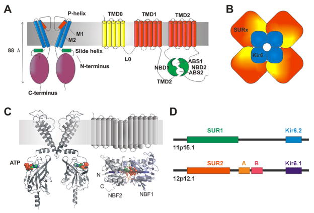
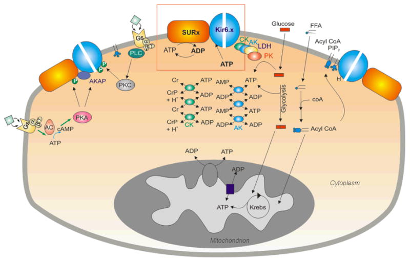
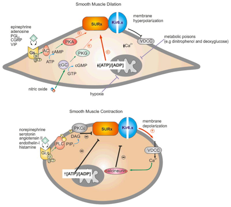
ATP-sensitive potassium (K(ATP)) channels are present in the surface and internal membranes of cardiac, skeletal, and smooth muscle cells and provide a unique feedback between muscle cell metabolism and electrical activity. In so doing, they can play an important role in the control of contractility, particularly when cellular energetics are compromised, protecting the tissue against calcium overload and fiber damage, but the cost of this protection may be enhanced arrhythmic activity. Generated as complexes of Kir6.1 or Kir6.2 pore-forming subunits with regulatory sulfonylurea receptor subunits, SUR1 or SUR2, the differential assembly of K(ATP) channels in different tissues gives rise to tissue-specific physiological and pharmacological regulation, and hence to the tissue-specific pharmacological control of contractility. The last 10 years have provided insights into the regulation and role of muscle K(ATP) channels, in large part driven by studies of mice in which the protein determinants of channel activity have been deleted or modified. As yet, few human diseases have been correlated with altered muscle K(ATP) activity, but genetically modified animals give important insights to likely pathological roles of aberrant channel activity in different muscle types.


Figures

Fig. 1
Fig. 1. The structural basis of KATP channel activity
(A) KATP channels are formed from Kir6 (left) and SUR (right) subunits. Kir6 consists of a tetrameric arrangement of subunits, each consisting of two transmembrane helioces (M1, M2) a pore-forming region (including the p-helix and selectivity fitler) and cytoplasmic N-temrinus (including the amphipathic slide helix) and C-terminus. SUR consists of the TM0 and L0 regions that interact with and mosulate gating of Kir6, followed by two additional 6-helix TM regions, each followed by a nucleotide binding fold (NBF). The two nucleotide binding folds come together to generate two nucleotide interacting sites at their interface. (B) The KATP channel is an octameric structure of Kir6 and SUR subunits. (C) Kir6 and SUR can be modeled on the structure of other prokaryotic and eukaryotic proteins. Such structures predict one ATP binding site at each of the Kir6 interfaces, and Mg-nucleotide binding sites in the dimeric NBF structures of SUR. (D) Human SUR and Kir6 gene structures indicate that each pair are located adjacent to each other on two chromosomes.
Fig. 2
Fig. 2. Complexities of KATP channel regulation in striated muscle
The balance of ATP synthesis and usage, reflected by ATP and ADP levels, is the major direct determinant of channel activity (red box). Metabolic enzymes, including adenylate kinase (AK), creatine kinase (CK), and lactate dehydrogenase (LDH) in the cytoplasm and physically associated with the channel may serve to amplify metabolic changes, or locally buffer and control ATP/ADP levels, thereby fine tuning channel activity. Non-nucleotide ligands, including PIP2, acyl-coA and H+ may also play a key role. PIP2 and acyl CoAs have powerful stimulatory effects that are antagonistic to ATP inhibition. In addition, hormone receptor activation can lead to protein phosphorylation (P) with both stimulatory and inhibitory effects on the channel. However, none of these molecules acts in isolation and the resultant KATP channel activity is an integrated response to a myriad of interrelated metabolic signals.
Fig. 3
Fig. 3. Signaling pathways involved in regulation and modulation of the smooth muscle KATP channel
The KATP channel determines, in part, the membrane potential of the smooth muscle cell and, hence, the contractile state. Vasoactive factors that inhibit KATP activity cause membrane depolarization, activation of voltage-dependent Ca2+-channels (VDCC), a rise in intracellular [Ca2+] and smooth muscle contraction. Conversely, factors that activate KATP prevent the depolarization-dependent rise in [Ca2+] and promote smooth muscle dilation. (A) Endogenous vasodilators, including epinephrine, adenosine, prostacyclin (PGl2), calcitonin-generated peptide (CGRP), and the vasoactive intestinal peptide (VIP), stimulate KATP activity through the classic G-protein (Gs)/adenylate cyclase (AC)/protein kinase A (PKA) signaling pathway. Serine/threonine phosphorylation at several key residue in SUR2B (Thr633, Ser1387, and Ser1465) is thought to underlie channel activation. Conversely, nitric oxide activates KATP through the protein kinase G (PKG) signaling pathway. Hypoxia and metabolic poisons (dinitrophenol and deoxygluxose) indirectly activate KATP by suppressing oxidative phosphorylation, resulting in a decrease in the ATP/ADP levels. The K+-channel opener drugs (KCOs) (eg. pinacidil and diazoxide) promote their anti-hypertensive effects by opening vascular smooth KATP channels via interaction with the SUR2B subunit. (B) Vasoconstrictor agonists that target VSM KATP to promote contraction include the neurotransmitter serotonin (5-HT), angiotensin-II, endothelin-I, and the pro-inflammatory histamine. Binding of vasoconstrictors to the G-protein receptor subtypes, Gi or Gq, indirectly activates the PKC-ε isoform. In turn, PKCε-mediated phosphorylation of a serine-rich motif in the C-terminus of Kir6.1 results in a decrease in the frequency of channel openings. Ca2+-activated calcineurin (phosphatase type 2B) inhibits VSM KATP although the mechanism is unclear. Sulfonylurea compounds (e.g glibenclamide) induce vasoconstriction by binding to the SUR2B subunit causing KATP channel closure.

References

    1. Aguilar-Bryan L, Nichols CG, Rajan AS, Parker C, Bryan J. Co-expression of sulfonylurea receptors and KATP channels in hamster insulinoma tumor (HIT) cells. Evidence for direct association of the receptor with the channel. J Biol Chem. 1992;267:14934–14940. - PubMed
    1. Aguilar-Bryan L, Nichols CG, Wechsler SW, Clement JPt, Boyd AE, 3rd, Gonzalez G, Herrera-Sosa H, Nguy K, Bryan J, Nelson DA. Cloning of the beta cell high-affinity sulfonylurea receptor: a regulator of insulin secretion. Science. 1995;268:423–426. - PubMed
    1. Allard B, Lazdunski M. Nucleotide diphosphates activate the ATP-sensitive potassium channel in mouse skeletal muscle. Pflugers Arch. 1992;422:185–192. - PubMed
    1. Allard B, Lazdunski M. Pharmacological properties of ATP-sensitive K+ channels in mammalian skeletal muscle cells. Eur J Pharmacol. 1993;236:419–426. - PubMed
    1. Allard B, Lazdunski M, Rougier O. Activation of ATP-dependent K+ channels by metabolic poisoning in adult mouse skeletal muscle: role of intracellular Mg(2+) and pH. J Physiol. 1995;485 (Pt 2):283–296. - PMC - PubMed

Publication types

MeSH terms