Review
. 2010 Jul;90(3):1165-94.
doi: 10.1152/physrev.00061.2009.

Hepatocyte death: a clear and present danger

Affiliations
Review

Hepatocyte death: a clear and present danger

Harmeet Malhi et al. Physiol Rev. 2010 Jul.

Abstract

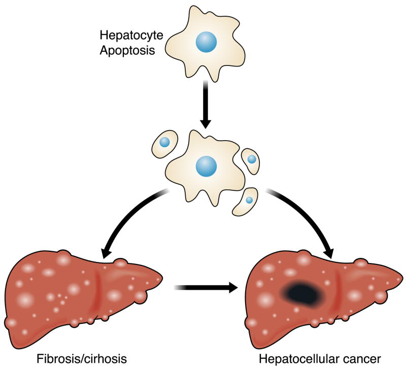
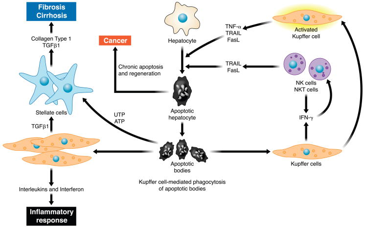
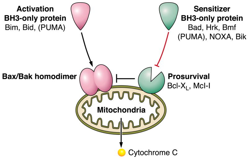
The hepatocyte is especially vulnerable to injury due to its central role in xenobiotic metabolism including drugs and alcohol, participation in lipid and fatty acid metabolism, its unique role in the enterohepatic circulation of bile acids, the widespread prevalence of hepatotropic viruses, and its existence within a milieu of innate immune responding cells. Apoptosis and necrosis are the most widely recognized forms of hepatocyte cell death. The hepatocyte displays many unique features regarding cell death by apoptosis. It is quite susceptible to death receptor-mediated injury, and its death receptor signaling pathways involve the mitochondrial pathway for efficient cell killing. Also, death receptors can trigger lysosomal disruption in hepatocytes which further promote cell and tissue injury. Interestingly, hepatocytes are protected from cell death by only two anti-apoptotic proteins, Bcl-x(L) and Mcl-1, which have nonredundant functions. Endoplasmic reticulum stress or the unfolded protein response contributes to hepatocyte cell death during alterations of lipid and fatty acid metabolism. Finally, the current information implicating RIP kinases in necrosis provides an approach to more fully address this mode of cell death in hepatocyte injury. All of these processes contributing to hepatocyte injury are discussed in the context of potential therapeutic strategies.


Figures

FIG. 1
FIG. 1
Hepatocyte apoptosis as a mechanism of liver injury and carcinogenesis. The precise mechanism(s) by which apoptosis promotes liver inflammation and fibrosis is unclear but well described in the literature. The cell turnover also provides a platform for cancer-initiating mutations, while the proapoptotic pressure is an impetus to develop mechanisms to avoid apoptosis (a hallmark of cancer).
FIG. 2
FIG. 2
Cellular mechanisms of hepatic injury and fibrosis due to hepatocyte apoptosis. Hepatocyte apoptosis results in the formation of apoptotic bodies. Engulfment of apoptotic bodies by liver resident macrophages or Kupffer cells enhances their expression of death ligands such as TRAIL, Fas, and tumor necrosis factor (TNF)-α. These death ligands in a feed-forward loop further promote hepatocyte apoptosis and generation of apoptotic bodies. Engulfment of apoptotic bodies by stellate cells promotes their activation and enhances their development into myofibroblasts, which secrete collagen type 1 and transforming growth factor (TGF)-β1, promoting development of fibrosis and cirrhosis. Recently, apoptotic cells were shown to release the nucleotides UTP and ATP, which bind P2Y2 purinergic receptors on myofibroblasts enhancing their secretion of collagen. The constant cell turnover and proapoptotic pressure are also permissive for the development of hepatocellular carcinoma.
FIG. 3
FIG. 3
Caspase structure. Caspases consist of a catalytic domain organized in a large subunit (~20 kDa), a small subunit (~10 kDa), and a prodomain of variable length. Effector caspases exhibit a short prodomain, whereas initiator caspases have long prodomains that direct binding to protein complexes. Caspase-directed cleavages at aspartate residues in a linker region between the large and the small subunit and between the prodomain and the large subunit generate the formation of the 20- and 10-kDa polypeptides that oligomerize to form the heterotetrameric active form of protease. The catalytic cysteine is embedded in the conserved pentapeptide QACXG motif within the large subunit.
FIG. 4
FIG. 4
Bcl-2 family proteins regulate the mitochondrial pathway of apoptosis. Mitochondrial dysfunction is induced by homodimerization of Bax or Bak within the outer mitochondrial membrane. The homodimerization of these proteins can be directly stimulated by the BH3-only proteins Bim or Bid, and perhaps PUMA; hence, these three proteins are referred to as direct activators. Bax and Bak homodimerization is also kept in check by pro-survival Bcl-2 proteins, which in the hepatocyte are Bcl-xL and Mcl-1. The pro-survival function of these proteins can be repressed by the sensitizer BH3-only proteins Bad, Hrk, Bmf, NOXA, Bik, and PUMA. BH3-only protein repression of Mcl-1 and Bcl-xL then allows activation of Bax and/or Bak by an as yet unclear mechanism.
FIG. 5
FIG. 5
Death receptor signaling and its interdigitation with the mitochondrial pathway of apoptosis. Ligation of a death receptor by its ligand results in the recruitment of pro-caspases 8 and 10 to a receptor complex via homotypic interactions with FADD (or the other adaptor protein TRADD). Within this complex, caspases 8 and 10 undergo auto-activation. Active caspase 8/10 can directly activate caspase 3, an executioner caspase (type I cells), or cleave Bid, a BH3-only member of the Bcl-2 family, to generate truncated Bid (tBid) which in turn induces mitochondrial dysfunction (type II cells). X-linked inhibitor of apoptosis (XIAP) determines which pathway results in cell death. In hepatocytes, activation of the mitochondrial pathway is necessary to overcome resistance by XIAP.
FIG. 6
FIG. 6
Danger associated molecular patterns (DAMPs) and acetaminophen-induced liver injury. DAMPs are intracellular signals released from dying cells, both necrotic and apoptotic cells. Nuclear and cytoplasmic DAMPs including the protein HMGB-1, nucleosides, uric acid, and heat shock proteins are released from dying cells. These DAMPs activate the innate immune system and initiate a secondary cascade of inflammation and injury. In the case of acetaminophen (AAP), animals models have elucidated a role for HMGB-1-initiated signaling events indicated in this figure with double asterisk (**). HMGB-1, in a TLR9-dependent manner, activates the inflammasome consisting of Nalp3 (NACHT, LRR, and pyrin domain-containing protein 3), ASC (apoptosis-associated speck-like protein containing CARD), and procaspase 1. Activation and autoproteolytic cleavage of procaspase 1 yields active caspase 1 in the inflammasome. Independent of the inflammasome, HMGB1-induced TLR9 activation results in enhanced expression of pro-interleukin (IL)-1β and pro-IL-18. Cleavage of pro-IL-1β and pro-IL-18 to active IL-1β and IL-18 is catalyzed by caspase 1. CD24 and Siglec-G are negative regulators of HMGB1-activated innate immune responses. HMGB-1 can bind LPS and enhance TLR4 activation, as well as activate TLR2; however, the contribution of these two pathways to AAP-induced liver injury has not yet been elucidated.
FIG. 7
FIG. 7
Toll-like receptor 4 (TLR4) signaling in Kupffer cells. Shown here is a Kupffer cell with cell surface TLR4 expression. Upon ligation with lipopolysaccharide, a process facilitated by the coreceptors CD14 and MD2 as well as LPS-binding protein, the proximal adaptor molecules MyD88 and TIRAP are recruited, activating kinase cascades that result in activation of NF-κB, and the MAPK ERK1, p38, and JNK. Subsequent TLR4 internalization and recruitment of the adaptor proteins TRIF and TRAM activate type 1 interferon production. Initial TLR4 activation can occur downstream of changes in gut permeability that permit greater translocation of bacterial LPS. TLR4 can also be activated by DAMPs, including HMGB-1 released from dying cells. Upon activation of Kupffer cells, many pro- and anti-inflammatory cytokines are produced, which serve a physiological as well as pathological role in the liver. Further recruitment of leukocytes with amplification of cytokine production occurs. Death ligands (FasL and TRAIL) activate death receptor-mediated hepatocyte apoptosis. Oxidant stress can also lead to cell death. DAMPs released from dying cells further accentuate TLR4 signaling. TGF-β produced by Kupffer cells promotes the activation of hepatic stellate cells, although multiple other signals also govern this complex process, including engulfment of apoptotic hepatocytes and other TLRs.
FIG. 8
FIG. 8
The bile acid-induced/death receptor-mediated apoptosis pathway. Hydrophobic bile acid stimulates Golgi-associated and microtubule-dependent Fas trafficking to plasma membrane, resulting in an increased density of cell surface Fas. In a FasL-independent manner, Fas undergoes oligomerization and recruits FADD, which, in turn, binds to pro-caspase 8 and facilitates its activation by autocatalytic processes. Active caspase 8 cleaves Bid, whose truncated form translocates to mitochondria and cooperates with Bax to induce MOMP. This results in release of apoptogenic factors, such as cytochrome c, AIF, and SMAC, into the cytosol, which ultimately promote the activation of effector caspases 3, 6, and 7 and cell death. At the same time, hydrophobic bile acids also cause upregulation of death receptor 5 (DR5) via JNK-mediated activation of the transcription factor Sp1 (specificity protein 1), sensitizing the hepatocytes to TRAIL-induced apoptosis.
FIG. 9
FIG. 9
TNF-R1 signaling pathways. Engagement of TNF-R1 by TNF-α induces recruitment of TRADD, RIP1, TRAF2, and c-IAP1/2 to the receptor to form the membrane-bound complex I. Two distinct pathways originate from the association of RIP1 and TRAF-2 to the receptor. The first one promotes survival by activation of the catalytic IKK complex, leading to phosphorylation of the NF-κB inhibitory protein IκBα and its degradation via the ubiquitin-proteasome pathway, allowing NF-κB to translocate to the nucleus and initiate transcription of anti-apoptotic target genes, such as cFLIP, cIAP-1, cIAP-2, TRAF1, TRAF2, XIAP, and Gadd45β. The second one leads to activation of JNK via the consequential activation of MEKK1/3 and MKK4/7. Activation of JNK is usually transient due to prompt inhibition by NF-κB-regulated proteins, such as XIAP and Gadd45β; however, sustained activation of JNK due to defects in NF-κB transcriptional activity results in c-FLIP degradation, enhanced caspase 8 activation, and apoptosis. Following the transient formation of complex I, the receptor is internalized by endocytosis and RIP1, TRAF2 and TRADD dissociated from TNF-R1, allowing the recruitment of FADD and caspase 8. The newly formed cytosolic complex (named complex II) promotes the activation of caspase 8, initiating the apoptotic cascade. This apoptotic pathway is inhibited by NF-κB-mediated expression of cFLIPL, which binds to caspase 8 and prevents its proteolytic activation.
FIG. 10
FIG. 10
The lysosomal pathway of apoptosis in hepatocytes. Death receptor engagement by their cognate ligands results in formation of a DISC promoting activation of caspase 8. Caspase 8 cleaves and activates Bid, which, in hepatocytes, facilitates lysosomal membrane permeabilization (LMP), likely by activating Bax. In cholangiocytes, another BH3-only protein, Bim, but not Bid, cooperates with Bax to induce LMP following phosphorylation/activation by JNK. LMP results in release of lysosomal components into the cytosol. Although not the only lysosomal protease released, cathepsin B plays a pivotal role in lysosomal-mediated cytotoxicity in liver cells by promoting mitochondrial dysfunction. The mechanisms by which cathepsin B causes MOMP are still largely unknown; however, cleavage and activation of Bid/Bax and caspase 2 have been described in different experimental models.
FIG. 11
FIG. 11
Unfolded protein response and cell death. Endoplasmic reticulum (ER) stress, such as the accumulation of misfolded proteins in the ER lumen, activates the unfolded protein response (UPR), aimed at restoring ER homeostasis through translational and transcriptional pathways. The UPR sensors IRE1α, PERK, and ATF6α activate these pathways. Activation of IRE1α leads to the unconventional splicing of the transcription factor XBP1 to the transcriptionally active spliced XBP1 (XBP1s), which activates UPR target genes aimed at restoration of ER homeostasis. Via the adaptor protein TRAF2, IRE1α can activate JNK. ATF6α is cleaved in the Golgi to an active transcription factor which induces expression of UPR target genes, also aimed at restoration of homeostasis. Phosphorylation of eIF2α by PERK results in attenuation of translation and selective translation of ATF4 and its target CHOP. Failure of restoration of ER homeostasis results in cell death. Some possible mechanisms include CHOP-induced expression of the proapoptotic protein Bim, and DR5, perturbations in ER calcium, and JNK-induced cell death. The anti-apoptotic protein Bax inhibitor 1 (BI-1) is a negative regulator of IRE1α, as well as negative regulator of ER-stress induced cell death.
FIG. 12
FIG. 12
Molecular mechanisms involved in hepatocyte lipoapoptosis. Saturated FFAs accumulate in the ER eliciting an ER stress response mediated by phosphorylation and activation of IRE1α and PERK. Phosphorylated PERK promotes induction of the transcription factor CHOP, which upregulates the expression of the pro-apoptotic BH3-only protein Bim. Phosphorylated IRE1α activates JNK, which, in turn, phosphorylates the transcription factor c-Jun. Activation of c-Jun results in transcriptional upregulation of the pro-apoptotic BH3-only protein PUMA. Bim and PUMA activate the pro-apoptotic executioner protein Bax, resulting in MOMP, activation of effector caspases, and apoptosis. In addition, CHOP upregulates the expression of death receptors, such as DR5, increasing the susceptibility of steatotic hepatocytes to circulating death ligands (e.g., TRAIL).

Similar articles

Cited by

References

    1. Adams LA, Lymp JF, St Sauver J, Sanderson SO, Lindor KD, Feldstein A, Angulo P. The natural history of nonalcoholic fatty liver disease: a population-based cohort study. Gastroenterology. 2005;129:113–121. - PubMed
    1. Ahlenstiel G, Titerence RH, Koh C, Edlich B, Feld JJ, Rotman Y, Ghany MG, Hoofnagle JH, Liang TJ, Heller T, Rehermann B. Natural killer cells are polarized toward cytotoxicity in chronic hepatitis C in an interferon-alpha-dependent manner. Gastroenterology. 2010;138:325–335. - PMC - PubMed
    1. Akazawa Y, Mott JL, Bronk SF, Werneburg NW, Kahraman A, Guicciardi ME, Meng XW, Kohno S, Shah VH, Kaufmann SH, McNiven MA, Gores GJ. Death receptor 5 internalization is required for lysosomal permeabilization by TRAIL in malignant liver cell lines. Gastroenterology. 2009;136:2365–2376. - PMC - PubMed
    1. Amaral JD, Viana RJ, Ramalho RM, Steer CJ, Rodrigues CM. Bile acids: regulation of apoptosis by ursodeoxycholic acid. J Lipid Res. 2009;50:1721–1734. - PMC - PubMed
    1. Araya J, Rodrigo R, Videla LA, Thielemann L, Orellana M, Pettinelli P, Poniachik J. Increase in long-chain polyunsaturated fatty acid n-6/n-3 ratio in relation to hepatic steatosis in patients with non-alcoholic fatty liver disease. Clin Sci. 2004;106:635–643. - PubMed

Publication types

Substances