. 2010 Oct;18(10):1857-64.
doi: 10.1038/mt.2010.155. Epub 2010 Jul 27.

Reactive bone marrow stromal cells attenuate systemic inflammation via sTNFR1

Affiliations

Reactive bone marrow stromal cells attenuate systemic inflammation via sTNFR1

Hiroshi Yagi et al. Mol Ther. 2010 Oct.

Abstract

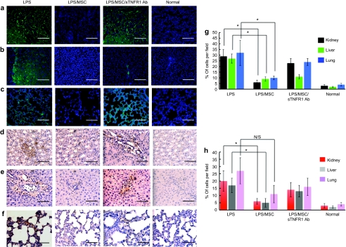
Excessive systemic inflammation following trauma, sepsis, or burn could lead to distant organ damage. The transplantation of bone marrow stromal cells or mesenchymal stem cells (MSCs) has been reported to be an effective treatment for several immune disorders by modulating the inflammatory response to injury. We hypothesized that MSCs can dynamically secrete systemic factors that can neutralize the activity of inflammatory cytokines. In this study, we showed that cocultured MSCs are able to decrease nuclear factor κ-B (NFκB) activation in target epithelial cells incubated in inflammatory serum conditions. Proteomic screening revealed a responsive secretion of soluble tumor necrosis factor (TNF) receptor 1 (sTNFR1) when MSCs were exposed to lipopolysaccharide (LPS)-stimulated rat serum. The responsive effect was eliminated when NFκB activation was blocked in MSCs. Intramuscular transplantation of MSCs in LPS-endotoxic rats decreased a panel of inflammatory cytokines and inflammatory infiltration of macrophages and neutrophils in lung, kidney, and liver when compared to controls. These results suggest that improvements of inflammatory responses in animal models after local transplantation of MSCs are at least, in part, explained by the NFκB-dependent secretion of sTNFR1 by MSCs.


Figures

Figure 1
Figure 1
Human mesenchymal stem cells (hMSCs) reduce NFκB activation in a reporter cell line. Fluorescence-activated cell sorting (FACS) analysis was performed to analyze GFP+ cell ratio in cocultures of hMSCs and the NFκB-GFP cell line (1:5 ratio) after 24 hours. (a) Different doses of TNFα (0, 1, 5, 10, 15, 20, 50, and 100 ng/ml) were used to validate the reporter cell line (NFκB-GFP). (b) Quantification of FACS analysis for GFP+ cells when cultured in inflammatory serum with or without hMSCs as well as no treatment as a control. Error bars represent mean ± SD (*P = 0.0003). (c) Results were corroborated by fluorescent microscopy. DAPI was used to counterstain nuclei. c, Bar = 100 µm. (d) Representative data were analyzed by flow cytometry. GFP, green fluorescent protein; LPS, lipopolysaccharide; TNF-α, tumor necrosis factor-α.
Figure 2
Figure 2
Characterization of soluble receptor secretion and inflammatory serum-responding secretion by human mesenchymal stem cells (hMSCs). (a) Human sTNFR1 and 2, sCD40, sCD40L, and sIL1R2 as well as IL10 were measured in the culture medium of 0.5 × 10⁶ of hMSCs after 24 hours of exposure to culture media containing 20% normal serum or serum derived from endotoxemic animals (*P = 0.0135). (b) hMSCs were exposed to 20% of serum harvested from healthy rats (normal serum) or 6 hours after rats were treated with LPS (LPS serum). sTNFR1 levels in each medium were analyzed after 2 hours with (c) control peptide and with (d) cell-permeable NFκB inhibitor peptide (*P = 0.0226, **P = 0.0114). N/S, no significant difference. Error bars represent mean ± SD. LPS, lipopolysaccharide.
Figure 3
Figure 3
Transplantation of mesenchymal stem cells (MSCs) ameliorates proinflammatory cytokines in endotoxemic animals. (a–c) Hematoxylin and eosin staining (a) and immunohistochemical analysis (b) ×40 and (c) ×20 were performed using CD105 for the detection of hMSCs in the muscle where MSCs (2 × 10⁶ cells) were transplanted 24 hours before. (d–f) CD105 staining was performed in three vital organs, namely, (d) kidney, (e) liver, and (f) lung from animals 24 hours after receiving hMSC transplantation intramuscularly. DAPI was used to counterstain nuclei. a–f, Bar = 100 µm. Magnifications are ×20. (g) Data represent human sTNFR1 levels in serum of rats exposed to intraperitoneal injection of LPS treated with or without MSC transplantation. Error bars represent mean ± SD (*P = 0.0135). (h) Cytokine levels in serum of rats exposed to intraperitoneal injection of LPS. Alterations of TNFα, IL6, and IFN-γ were observed in animals with intramuscular transplantation of hMSCs. Data representative of three independent trials with a total of N = 5 per group. Serum was analyzed for cytokines at 0, 6, 12, 24, and 48 hours after treatment by enzyme-linked immunosorbent assay. *P < 0.05: between serum from control animals and animals that were transplanted hMSCs. LPS, lipopolysaccharide; TNF-α, tumor necrosis factor-α.
Figure 4
Figure 4
Histological appearance of mesenchymal stem cell (MSC)–treated animals after LPS injection. (a–c) Myeloperoxidase staining for detecting neutrophil infiltration of three vital organs; (a) kidney, (b) liver, and (c) lung, and (d–f) CD163 staining for macrophage infiltration of three vital organs; (d) kidney, (e) liver, and (f) lung were performed on the tissues from MSC transplanted animals 24 hours after LPS injection, whereas the samples from animals without cell transplantation after LPS, animals with cell transplantation treated with sTNFR1 antibody, and normal animals were served as controls. DAPI was used to counterstain nuclei in picture a–c. Magnifications are ×40. (g) Neutrophil infiltration of three vital organs in hMSC-treated rats was evaluated comparing to tissues from nontreated rats with LPS intoxication based on myeloperoxidase staining by the cell count in a field of ×40. (h) Macrophage infiltration of three vital organs in hMSC-treated rats was evaluated comparing to tissues from nontreated rats with LPS intoxication based on CD163 staining by the cell count in a field of ×40 (*P < 0.05). N/S, no significant difference. Error bars represent mean ± SD. a–f, Bar = 100 µm. LPS, lipopolysaccharide.

Similar articles

Cited by

References

    1. Heron M, Hoyert DL, Murphy SL, Xu J, Kochanek KD., and, Tejada-Vera B. Deaths: final data for 2006. Natl Vital Stat Rep. 2009;57:1–134. - PubMed
    1. Robertson CM., and, Coopersmith CM. The systemic inflammatory response syndrome. Microbes Infect. 2006;8:1382–1389. - PubMed
    1. Heidecke CD, Hensler T, Weighardt H, Zantl N, Wagner H, Siewert JR, et al. Selective defects of T lymphocyte function in patients with lethal intraabdominal infection. Am J Surg. 1999;178:288–292. - PubMed
    1. Tracey KJ, Fong Y, Hesse DG, Manogue KR, Lee AT, Kuo GC, et al. Anti-cachectin/TNF monoclonal antibodies prevent septic shock during lethal bacteraemia. Nature. 1987;330:662–664. - PubMed
    1. Deans KJ, Haley M, Natanson C, Eichacker PQ., and, Minneci PC. Novel therapies for sepsis: a review. J Trauma. 2005;58:867–874. - PubMed

Publication types

MeSH terms

Substances

LinkOut - more resources