. 2010 Nov;3(6):622-32.
doi: 10.1038/mi.2010.39. Epub 2010 Jul 21.

Intestinal epithelia activate anti-viral signaling via intracellular sensing of rotavirus structural components

Affiliations

Intestinal epithelia activate anti-viral signaling via intracellular sensing of rotavirus structural components

A H Frias et al. Mucosal Immunol. 2010 Nov.

Abstract

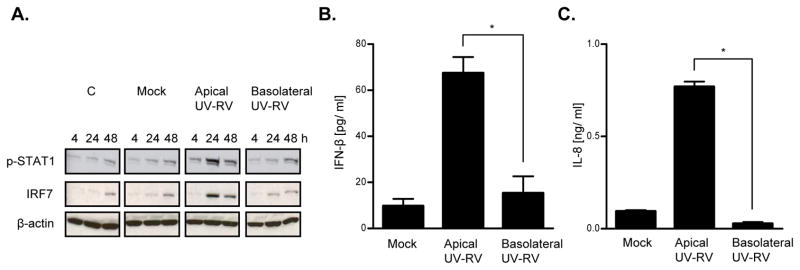
Rotavirus (RV), a leading cause of severe diarrhea, primarily infects intestinal epithelial cells (IECs) causing self-limiting illness. To better understand innate immunity to RV, we sought to define the extent to which IEC activation of anti-viral responses required viral replication or could be recapitulated by inactivated RV or its components. Using model human intestinal epithelia, we observed that RV-induced activation of signaling events and gene expression typically associated with viral infection was largely mimicked by administration of ultraviolet (UV)-inactivated RV. Use of anti-interferon (IFN) neutralizing antibodies revealed that such replication-independent anti-viral gene expression required type I IFN signaling. In contrast, RV-induction of nuclear factor-κB-mediated interleukin-8 expression was dependent on viral replication. The anti-viral gene expression induced by UV-RV was not significantly recapitulated by RV RNA or RV virus-like particles although the latter could enter IEC. Together, these results suggest that RV proteins mediate viral entry into epithelial cells leading to intracellular detection of RV RNA that generates an anti-viral response.


Conflict of interest statement

DISCLOSURE

The authors declared no conflicts of interest.

Figures

Figure 1
Figure 1. Anti-viral protein expression in RV-infected intestinal epithelia
Human intestinal epithelia (HT29) were grown to confluence in 6 well plates (A, B, C) or collagen-coated permeable supports (D, E, F) and infected with RV (MOI 0.5–1). Control samples were treated with trypsin diluted in SFM (Mock) or SFM alone (C). Cell lysates and supernatants were collected 0–48 hours post-inoculation (hpi). Western blot analyses were performed to assess viral protein (VP6) synthesis and protein expression of the indicated anti-viral markers in cell lysates (A, D). ELISA assays were used to measure IFN-β (B, E) and IL-8 (C, F) secretion in supernatants over time. Data in A, B, D, and E are results of a single experiment and representative of 3 separate experiments that gave similar results. Data in C and F reflects the mean +/− standard error of the mean (SEM) of 3 parallel experiments. Statistically significant differences P< 0.05 are denoted as starred values (*).
Figure 2
Figure 2. Anti-viral protein expression exhibited in RV-infected and UV-RV stimulated epithelia
Confluent intestinal epithelia (HT29) were grown in 6 well plates and treated with RV and UV-RV (MOI 1). Control samples were exposed to trypsin diluted in SFM (Mock), irradiated cellular debris from a mock preparation of UV-RV (Mock Irradiation), or SFM alone (C). Cell lysates and supernatants were collected at various time points (0–48 hpi). Western blot analyses were performed to assess viral protein (VP6) synthesis and protein expression of the indicated anti-viral markers in cell lysates (A, D). ELISA assays were used to measure secretion of IFN-β (B) and IL-8 (C) in supernatants at 48 hpi. Data in A, B and D are results of a single experiment and representative of 3 separate experiments that gave similar results. Data in C is the mean +/− SEM of 3 parallel experiments. Statistically significant differences P< 0.05 are denoted as starred values (*).
Figure 3
Figure 3. Anti-viral protein expression exhibited in epithelia treated with RV and UV-RV in the presence or absence of trypsin
Intestinal epithelial monolayers (HT29) grown in 6 well plates were treated with RV and UV-RV (MOI 1) in the presence or absence of trypsin (24 hpi). Control samples received equivalent amounts of trypsin diluted in SFM (Mock) or SFM alone. Trypsin and trypsin-free treatments are denoted as (+) and (−) symbols, respectively. Western blot analysis was used to detect anti-viral gene expression in cell lysates (A). ELISA assays were performed to measure secretion of IFN-β (B) and IL-8 (C) in supernatants at 24 hpi. Data in A shows results of a single experiment and is representative of 3 separate experiments that gave similar results. Data in B and C is the mean +/− SEM of 3 parallel experiments. Statistically significant differences P< 0.05 are denoted as starred values (*).
Figure 4
Figure 4. Anti-viral protein expression exhibited in epithelia treated apically and basolaterally with UV-RV
Intestinal epithelial monolayers (HT29) were grown on collagen-coated permeable supports and infected either apically or basolaterally with UV-RV (MOI 1). Control samples received equivalent amounts of trypsin diluted in SFM (Mock) or SFM alone (C). Cell lysates and supernatants were collected at various time points (0–48 hpi). Western blot analyses were performed to assess protein expression of the indicated anti-viral markers in cell lysates (A). ELISA assays were used to measure IFN-β (B) and IL-8 (C) secretion in supernatants at 48 hpi. Data in A shows results of a single experiment and is representative of 3 separate experiments that gave similar results. Data in B and C reflects the mean +/− SEM of 3 parallel experiments. Statistically significant differences P< 0.05 are denoted as starred values (*).
Figure 5
Figure 5. Transcription profiles of epithelia stimulated with RV, UV-RV, and RV in the presence of Type 1 IFN (α/β) antibodies
Intestinal epithelial (HT29) cell monolayers were grown in 6 well plates and infected with RV and UV-RV (MOI 1), and RV (MOI 1) plus Type 1 IFN antibodies (anti-IFN α/β). Control samples received equivalent amounts of trypsin diluted in SFM (Mock). Experiments were performed in biological triplicates. At 24 hpi, cell lysates were extracted for RNA and microarray analyses were performed to assess global transcription of genes. Heat map illustration of genes induced by RV and UV-RV with > 1.3 fold change relative to mock (A). qRT-PCR results used to confirm mRNA synthesis of select anti-viral genes at 24 hpi (B, C, D, E). Data in A shows results of 3 parallel experiments. Data in B, C, D, and E reflects the mean +/− SEM of 3 parallel experiments. Statistically significant differences P< 0.05 are denoted as starred values (*).
Figure 6
Figure 6. Anti-viral protein expression exhibited in epithelia treated with UV-RV, RV VLPs and RV RNA
Confluent intestinal epithelia (HT29) were grown in 6 well plates and treated with UV-RV (MOI 0.5–1), RV RNA (0.5–5 μg/ml), and VLPs (0.5–5 μg/ml). Control samples received equivalent amounts of trypsin diluted in SFM (Mock). Cell lysates and supernatants were collected at various time points (0–24 hpi). Western blot analyses were used to detect anti-viral gene expression in cell lysates (A). ELISA assays were performed to measure IFN-β (B) or IL-8 (C) secretion in supernatants at 24 hpi. qRT-PCR analyses were utilized to confirm mRNA synthesis of select anti-viral genes from microarray experiments (see Table III). Data in A are results of a single experiment and representative of 3 separate experiments that gave similar results. Data in B, C, D, and E shows the mean +/− SEM of 3 parallel experiments. Statistically significant differences P< 0.05 are denoted as starred values (*).
Figure 7
Figure 7. RV, UV-RV, and VLP cell entry during early stages of infection
Intestinal epithelial monolayers (HT29) were grown on collagen-coated permeable supports and treated apically with RV and UV-RV (MOI 10), and an amount of VLPs roughly equivalent to the estimated protein concentration in the RV preparation. Control samples were apically treated with an equivalent amount of trypsin in SFM (Mock) for 4 hpi. At 1 and 4 hpi, cells were fixed, stained and examined via confocal fluorescence microscopy for presence of rotaviral proteins (green) and F-actin (red) in the sub-apical region of the cells (3 μm below the apical surface). Sub-apical images of cells stimulated with RV, UV-RV, and RV VLPs at 1 hpi, magnification 60X (A). Sub-apical images of cells treated with RV and UV-RV at 1 and 4 hpi, magnification 40X (B). Data in A and B are results of a single experiment and representative of 3 separate experiments. Scale reflects distance of 10 μm.

Similar articles

Cited by

References

    1. Glass RI, Parashar UD, Bresee JS, Turcios R, Fischer TK, Widdowson MA, et al. Rotavirus vaccines: current prospects and future challenges. Lancet. 2006 Jul 22;368(9532):323–32. - PubMed
    1. Parashar UD, Hummelman EG, Bresee JS, Miller MA, Glass RI. Global illness and deaths caused by rotavirus disease in children. Emerg Infect Dis. 2003 May;9(5):565–72. - PMC - PubMed
    1. Anderson EJ, Weber SG. Rotavirus infection in adults. Lancet Infect Dis. 2004 Feb;4(2):91–9. - PMC - PubMed
    1. Greenberg HB, Estes MK. Rotaviruses: from pathogenesis to vaccination. Gastroenterology. 2009 May;136(6):1939–51. - PMC - PubMed
    1. Jayaram H, Estes MK, Prasad BV. Emerging themes in rotavirus cell entry, genome organization, transcription and replication. Virus Res. 2004 Apr;101(1):67–81. - PubMed

Publication types

MeSH terms