

doi: 10.1364/OL.35.000043.
Rapid volumetric angiography of cortical microvasculature with optical coherence tomography
Affiliations
- PMID: 20664667
- PMCID: PMC2912612
- DOI: 10.1364/OL.35.000043
Item in Clipboard
Rapid volumetric angiography of cortical microvasculature with optical coherence tomography
Opt Lett.
.
Abstract
We describe methods and algorithms for rapid volumetric imaging of cortical vasculature with optical coherence tomography (OCT). By optimizing system design, scanning protocols, and algorithms for visualization of capillary flow, comprehensive imaging of the surface pial vasculature and capillary bed is performed in approximately 12 s. By imaging during hypercapnia and comparing with simultaneous CCD imaging, the sources of contrast of OCT angiography are investigated.
Figures
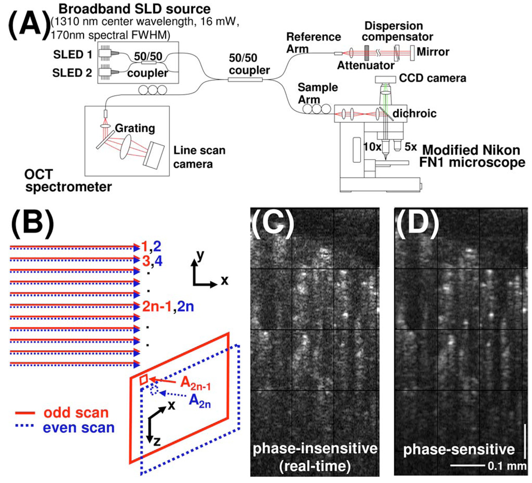
(Color online) (A) OCT system and microscope schematic. (B) Three-dimensional scan protocol for performing OCT angiography that samples each transverse location twice. (C),(D) Examples of cross-sectional OCT angiograms generated by phase-insensitive and phase-sensitive methods described in the text.

(Color online) (A) CCD image and (B) wide-field OCT angiogram of cranial window with (C) enlargement showing microvasculature. (D) 3D oblique slices and cross-sectional image (inset) showing distinct backscattering pattern due to shear-induced orientation of RBCs.
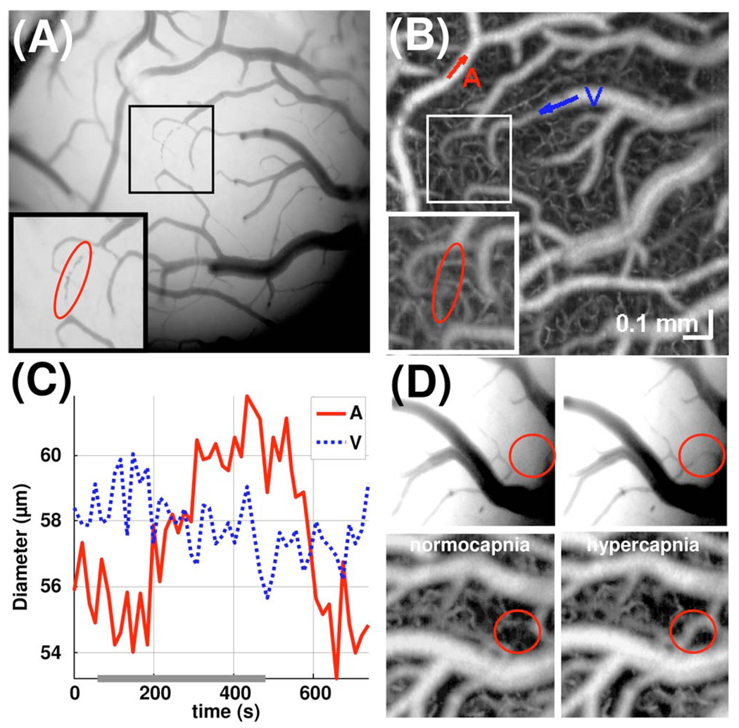
(Color online) (A) CCD image and (B) OCT angiogram of cortical vasculature during normocapnia. (C) Changes in vessel tone in representative artery A and vein V during 7.5% hypercapnia, shown as a line along the time axis. (D) Transient perfusion of arterial anastomosis during hypercapnia visualized by CCD imaging (above) and confirmed by OCT (below).

(Color online) (A), (C), Media 1: OCT imaging of the capillary bed during normocapnia. (B), (D), Media 2: hypercapnia, demonstrating flow redistribution (arrows).
References
Publication types
MeSH terms
Grants and funding
LinkOut - more resources
Full Text Sources
Other Literature Sources
