. 2010 Jul 7:16:1241-52.

NPR-B natriuretic peptide receptors in human corneal epithelium: mRNA, immunohistochemistochemical, protein, and biochemical pharmacology studies

Affiliations

NPR-B natriuretic peptide receptors in human corneal epithelium: mRNA, immunohistochemistochemical, protein, and biochemical pharmacology studies

Parvaneh Katoli et al. Mol Vis. .

Abstract

Purpose: To demonstrate the presence of natriuretic peptide receptors (NPRs) in primary human corneal epithelial cells (p-CEPI), SV40-immortalized CEPI cells (CEPI-17-CL4) and in human corneal epithelium, and to define the pharmacology of natriuretic peptide (NP)-induced cGMP accumulation.

Methods: NPR presence was shown by RT-PCR, western blot analysis, and indirect immunofluoresence. cGMP accumulation was determined using an enzyme immunoassay.

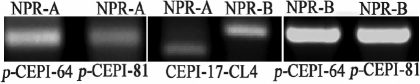
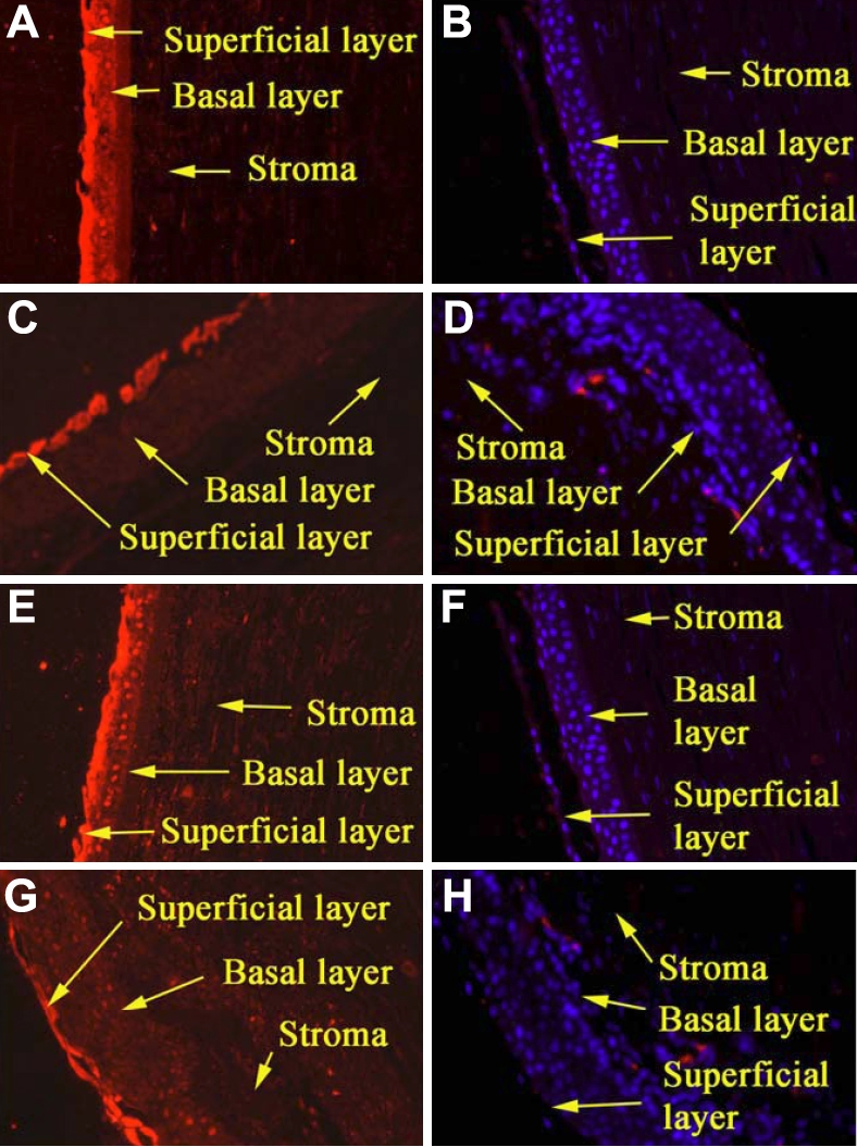
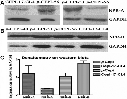
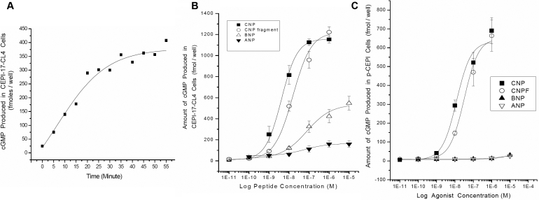
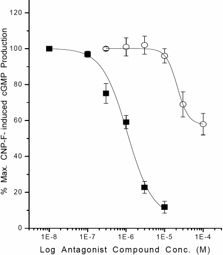
Results: p-CEPI and CEPI-17-CL4 cells expressed mRNAs for NPR-A and NPR-B. Proteins for both NPRs were present in these cells and in human corneal epithelium. C-type NP (CNP), atrial NP (ANP) and brain NP (BNP) stimulated the accumulation of cGMP in a concentration-dependent manner in p-CEPI cells (potency; EC(50s)): CNP (1-53 amino acids) EC(50)=24+/-5 nM; CNP fragment (32-53 amino acids) EC(50)=51+/-8 nM; ANP (1-28 amino acids) EC(50)=>10 microM; BNP (32 amino acids) EC(50)>10 microM (all n=3-4). While the NPs were generally more potent in the CEPI-17-CL4 cells than in p-CEPI cells (n=4-9; p<0.01), the rank order of potency of the peptides was essentially the same in both cell types. Effects of CNP fragment in p-CEPI and CEPI-17-CL4 cells were potently blocked by HS-142-1, an NPR-B receptor subtype-selective antagonist (K(i)=0.25+/-0.05 microM in CEPI-CL4-17; K(i)=0.44+/-0.09 microM in p-CEPIs; n=6-7) but less so by an NPR-A receptor antagonist, isatin (K(i)=5.3-7.8 microM, n=3-7).

Conclusions: Our studies showed the presence of NPR-A and NPR-B (mRNAs and protein) in p-CEPI and CEPI-17-CL4 cells and in human corneal epithelial tissue. However, detailed pharmacological studies revealed NPR-B to be the predominant functionally active receptor in both cell-types whose activation leads to the generation of cGMP. While the physiologic role(s) of the NP system in corneal function remains to be delineated, our multidisciplinary findings pave the way for such future investigations.


Figures

Figure 1
Figure 1
Detection of NPR-A and NPR-B mRNAs in human p-CEPI and CEPI-17-CL4 cells by RT–PCR. Lanes 1 and 2: NPR-A in human p-CEPI cells (donor ages 64 and 81); Lane 3: NPR-A in human CEPI-17-CL4 cells. The PCR product for NPR-A is equal to 179 bp. Lane 4: NPR-B in human CEPI-17-CL4 cells. The PCR product for NPR-B is equal to 237 bp. Lanes 5 and 6: NPR-B in human p-CEPI cells (donor ages 64 and 81).
Figure 2
Figure 2
Expression of NPR-A and NPR-B in paraffin sections of formalin-fixed human corneas as determined by indirect immunofluorescence. A, B: The presence of NPR-A in the epithelium of the central cornea. A shows stronger expression in the superficial layers when compared with the control (B). C, D: C shows the presence of NPR-A primarily in the superficial layers of the limbal epithelium. The control shows no expression (D). E, F: E shows strong expression of NPR-B primarily in the superficial cell layers of the central cornea compared when compared with the control (F). G, H: G shows that NPR-B is very distinctly confined to the superficial epithelium of the limbus compared with the control (H). All panels are at 10× magnification.
Figure 3
Figure 3
The expression of NPR-A and NPR-B in formalin-fixed human p-CEPI and CEPI-17-CL4 cells by indirect immunofluorescence is shown. A shows a strong expression of NPR-A in human p-CEPI cells, while B shows expression of NPR-A in CEPI-17-CL4 cells. C shows expression of NPR-B in human p-CEPI cells while D shows expression of NPR-B in CEPI-17-CL4 cells. E and F are DAPI labeled controls (treated with secondary antibody only) for human p-CEPI and CEPI-17-CL4 cells, respectively. Each panel is at 40× magnification.
Figure 4
Figure 4
Expression of NPR-A and NPR-B in human p-CEPI cells and CEPI-17-CL4 cells as determined by western blot analysis. A: The presence of NPR-A protein (band observed at 40–55 kDa) in CEPI-17-CL4 and in human p-CEPI cells (from 56, 53, and 56 year old donors) is shown. B: The presence of NPR-B protein (band observed at 24 kDa) is shown for three different donors of human p-CEPI cells (ages 40, 53, and 56) cells and in CEPI-17-CL4 cells. C: The expression of NPR-A and NPR-B in human p-CEPI and CEPI-17-CL4 cells was normalized to GAPDH. The figure shows an apparent lower expression of NPR-A in CEPI-17-CL4 cells than in human p-CEPI cells (p<0.1); the expression of NPR-B was higher in CEPI-17-CL4 than that in human p-CEPI cells (p<0.05). The NPR-B expression was greater than NPR-A expression in CEPI-17-CL4 cells (p<0.05). However, the both receptor subtypes were expressed to the same extent in the p-CEPI cells (p<0.1).
Figure 5
Figure 5
A: Time-course of increase in cGMP induced by CNP fragment in CEPI-17-CL4 cells is shown. Cyclic GMP concentration was measured after the cells were treated with CNP fragment for the indicated period. Each symbol represents a single datum point determined in duplicate. B and C: Effects of NPs on cGMP production in human p-CEPI and CEPI-17-CL4 cells are shown. Cyclic GMP concentration was measured after the cells were treated with the indicated concentrations of various NPs for 15 min. Each symbol represents a single datum point determined in duplicate. Results were obtained in 4 independent studies for CEPI-17-CL4 cells (B) and 3 independent studies for human p-CEPI cells (C).
Figure 6
Figure 6
Inhibition of CNP fragment-induced cGMP production in CEPI-17-CL4 cells by two NP receptor antagonists is shown. The effects of HS-142–1 (10 nM to 10 µM; closed square symbols) and isatin (0.3–100 µM; open circle symbols) on CNP fragment (30 nM)-induced cGMP generation were determined as described in the methods section. Data are mean±SEM from 6 to 7 experiments for HS-142–1, and from 7 experiments for isatin. Similar results were obtained using p-CEPI cells. The antagonist potencies of the two compounds in the two cell-types are provided in the Results section.

References

    1. deBold AJ. Atrial natriuretic factor: a hormone produced by the heart. Science. 1985;230:767–70. - PubMed
    1. Sudoh T, Kangawa K, Minamino N, Matsuo H. A new natriuretic peptide in porcine brain. Nature. 1988;332:78–81. - PubMed
    1. Sudoh T, Minamino N, Kangawa K, Matsuo H. C-type natriuretic peptide (CNP): a new member of natriuretic peptide family identified in porcine brain. Biochem Biophys Res Commun. 1990;168:863–70. - PubMed
    1. Drewett JG, Garbers DL. The family of guanylyl cyclase receptors and their ligands. Endocr Rev. 1994;15:135–62. - PubMed
    1. Potter LR, Yoder AR, Flora DR, Antos LK, Dickey DM. Natriuretic peptides: their structures, receptors, physiological functions and therapeutic applications. Handbook Exp Pharmacol. 2009;191:341–66. - PMC - PubMed

Publication types

Substances

LinkOut - more resources