. 2009 Aug 1;31(4):2-6.

Under the lash: Demodex mites in human diseases

Affiliations

Under the lash: Demodex mites in human diseases

Noreen Lacey et al. Biochem (Lond). .

Abstract

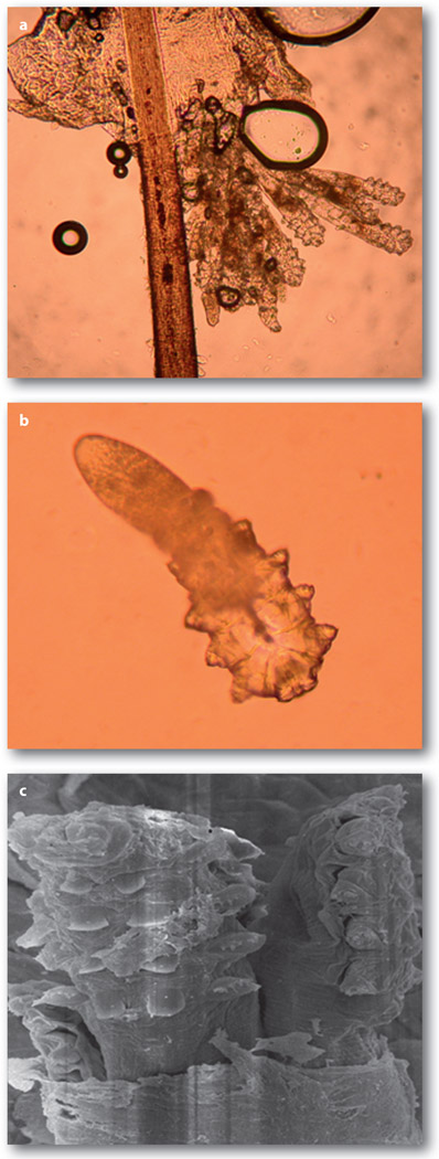
Demodex mites, class Arachnida and subclass Acarina, are elongated mites with clear cephalothorax and abdomens, the former with four pairs of legs. There are more than 100 species of Demodex mite, many of which are obligatory commensals of the pilosebaceous unit of mammals including cats, dogs, sheep, cattle, pigs, goats, deer, bats, hamsters, rats and mice. Among them, Demodex canis, which is found ubiquitously in dogs, is the most documented and investigated. In excessive numbers D. canis causes the inflammatory disease termed demodicosis (demodectic mange, follicular mange or red mange), which is more common in purebred dogs and has a hereditary predisposition in breeding kennels1. Two distinct Demodex species have been confirmed as the most common ectoparasite in man. The larger Demodex folliculorum, about 0.3-0.4 mm long, is primarily found as a cluster in the hair follicle (Figure 1a), while the smaller Demodex brevis, about 0.2-0.3 mm long with a spindle shape and stubby legs, resides solitarily in the sebaceous gland (Figure 1b). These two species are also ubiquitously found in all human races without gender preference. The pathogenic role of Demodex mites in veterinary medicine is not as greatly disputed as in human diseases. In this article, we review the key literature and our joint research experience regarding the pathogenic potential of these two mites in causing inflammatory diseases of human skin and eye. We hope that the evidence summarized herein will invite readers to take a different look at the life of Demodex mites in several common human diseases.


Figures

Figure 1
Figure 1
Microscopic features of adult D. folliculorum(a) and D. brevis (b). D. folliculorum tends to gather as a group in the follicle area of the hair or lash (a). In contrast, D. brevis tends to be in solitude and reside in the sebaceous gland. As a result, D. brevis is not readily detected during sampling of the lash. Under scanning electron microscopy, several D. folliculorum mites are together next to the hair follicle
Figure 2
Figure 2
Clinical features of Demodex infestation in the eye. The most common finding is CD on the skin surface where lashes emit (a). As mites reside close to the follicle, the orientation of lashes become disorganized (a), can turn in to touch the ocular surface (trichiasis) (b), and in the extreme cases results in the loss of lashes (madarosis) (c). Besides lashes, meibomian glands can be involved to manifest plugging of the orifices (d). The inflammation derived from lashes or meibomian glands of the lid margin can be spill over to the conjunctiva (e) and cause a number of corneal lesions, including nodular scar (f)
Figure 3
Figure 3
Unmasking embedded mites in CD by fluorescein solution. A representative lash with CD (a), in which embedded mites are revealed only after application of a drop of an aqueous solution containing fluorescein (b). An arrow marks the hidden mites (a) that are revealed by fluorescein drops. Note that this procedure of dissolving CD frequently generates air bubbles, and the change of colour is due to the presence of fluorescein
Figure 4
Figure 4
Migration of Demodex by lid scrub with TTO. In this eye with diffuse CD found in the lashes before treatment (a), lash to be epilated (marked by arrow) showed a fragment of CD attached to the lash and abundant Demodex embedded close to the lash follicle (d). After lid scrub in the orifice with 50% TTO, the lashes became clean and totally free of CD, but tails of Demodex were protruding from the lash roots (b, arrow). At 3 minutes after lid scrub, free Demodex was found on the trunk close to the skin surface, i.e. away from the lash follicle in the epilated lash (e). Rotating these lashes (shown in b) before epilation allowed us to detect a group of Demodex migrating along the lash trunk (f and g). If no lid scrub was carried out at home for one week, CD returned to the lashes 1 week later (c). With permission from Br. J. Ophthalmol.

References

    1. Baima B, Sticherling M. Acta Derm. Venereol. 2002;82:3–6. - PubMed
    1. Rufli T, Mumcuoglu Y. Dermatologica. 1981;162:1–11. - PubMed
    1. Pena GP, Andrade Filho JS. Rev. Inst. Med. Trop. Sao Paulo. 2000;42:171–173. - PubMed
    1. Forton F, Seys B. Br. J. Dermatol. 1993;128:650–659. - PubMed
    1. Norn MS. Dan. Med. Bull. 1971;18:14–17. - PubMed

LinkOut - more resources