. 2010 Mar 30;3(4):994-1015.
doi: 10.3390/ph3040994.

Antiproliferative Properties of Type I and Type II Interferon

Affiliations

Antiproliferative Properties of Type I and Type II Interferon

Joseph Bekisz et al. Pharmaceuticals (Basel). .

Abstract

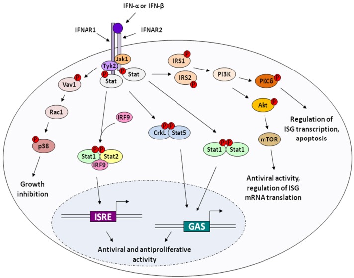
The clinical possibilities of interferon (IFN) became apparent with early studies demonstrating that it was capable of inhibiting tumor cells in culture and in vivo using animal models. IFN gained the distinction of being the first recombinant cytokine to be licensed in the USA for the treatment of a malignancy in 1986, with the approval of IFN-α2a (Hoffman-La Roche) and IFN-α2b (Schering-Plough) for the treatment of Hairy Cell Leukemia. In addition to this application, other approved antitumor applications for IFN-α2a are AIDS-related Kaposi's Sarcoma and Chronic Myelogenous Leukemia (CML) and other approved antitumor applications for IFN-α2b are Malignant Melanoma, Follicular Lymphoma, and AIDS-related Kapoisi's Sarcoma. In the ensuing years, a considerable number of studies have been conducted to establish the mechanisms of the induction and action of IFN's anti-tumor activity. These include identifying the role of Interferon Regulatory Factor 9 (IRF9) as a key factor in eliciting the antiproliferative effects of IFN-α as well as identifying genes induced by IFN that are involved in recognition of tumor cells. Recent studies also show that IFN-activated human monocytes can be used to achieve >95% eradication of select tumor cells. The signaling pathways by which IFN induces apoptosis can vary. IFN treatment induces the tumor suppressor gene p53, which plays a role in apoptosis for some tumors, but it is not essential for the apoptotic response. IFN-α also activates phosphatidylinositol 3-kinase (PI3K), which is associated with cell survival. Downstream of PI3K is the mammalian target of rapamycin (mTOR) which, in conjunction with PI3K, may act in signaling induced by growth factors after IFN treatment. This paper will explore the mechanisms by which IFN acts to elicit its antiproliferative effects and more closely examine the clinical applications for the anti-tumor potential of IFN.


Figures

Figure 1
Figure 1
Type I IFN Signaling. Type-I IFNs bind to a heterodimer composed of the transmembrane receptor subunits IFNAR1 and IFNAR2, which results in activation of the receptor-associated kinases Jak1 and Tyk2. In the canonical signaling pathway, the cytoplasmic signal transducers and activator of transcription (Stat) proteins are recruited to the receptor docking sites, phosphorylated, and dimerize to form the active transcription factors ISGF3 (Interferon stimulated gene factor 3), composed of phosphorylated Stat 1 and Stat2, and IRF9 (p48/ISGF3γ), and AAF/GAF (alpha activation factor/gamma activation factor), which is composed of two phosphorylated subunits of Stat1. These induce transcription of hundreds of interferon-stimulated genes (ISGs). IFN treatment also leads to induction of other non-canonical signaling pathways, including those involving p38 MAPK, Akt, and Crk. Upstream signaling from the IFNR complex leads to p38 phosphorylation, which modulates IFN activity and leads to growth inhibition of cells. CrkL and CrkII are tyrosine phosphorylated by Tyk2 after IFN treatment, and CrkL can also form transcription factor complexes with phosphorylated Stat5. Signaling pathways downstream of PI3K, involving Akt and mTOR, or PKCδ, are also important in mediating the biological activities of IFN.
Figure 2
Figure 2
Apoptotic Signaling in Response to DNA Damage. The caspase cascade is initiated by chemical damage to DNA which stimulates Bid cleavage leading to permeability transition of the mitochondrial membrane. The mitochondria releases cytochrome c in response to apoptotic signals and serves to activate Apaf-1 with consequent activation of Caspase 9 and the remainder of the caspase cascade. These caspases transmit the apoptotic signal which eventually leads to cell death.

References

    1. Isaacs A., Lindenmann J. Virus interference. I. The interferon. Proc. R. Soc. Lond B.Biol. Sci. 1957;147:258–267. doi: 10.1098/rspb.1957.0048. - DOI - PubMed
    1. Paucker K., Cantell K., Henle W. Quantitative studies on viral interference in suspended L cells : III. Effect of interfering viruses and interferon on the growth rate of cells. Virology. 1962;17:324–334. doi: 10.1016/0042-6822(62)90123-X. - DOI - PubMed
    1. Oxman M.N., Baron S., Black P.H., Takemoto K.K., Habel K., Rowe W.P. The effect of interferon on SV-40 T antigen production in SV-40-transformed cells. Virology. 1967;32:122–127. doi: 10.1016/0042-6822(67)90260-7. - DOI - PubMed
    1. Gresser I. Interferon and cancer: therapeutic prospects. Rev. Eur. Etud. Clin. Biol. 1970;15:23–27. - PubMed
    1. Sarma P.S., Shiu G., Baron S., Huebner R.J. Inhibitory effect of interferon on murine sarcoma and leukaemia virus infection in vitro. Nature. 1969;223:845–846. doi: 10.1038/223845a0. - DOI - PubMed

LinkOut - more resources