Multicenter Study
. 2010 Sep;457(3):309-17.
doi: 10.1007/s00428-010-0917-5. Epub 2010 Jul 28.

Multicentre validation study of nucleic acids extraction from FFPE tissues

Affiliations
Multicenter Study

Multicentre validation study of nucleic acids extraction from FFPE tissues

Serena Bonin et al. Virchows Arch. 2010 Sep.

Abstract

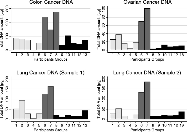
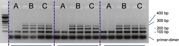
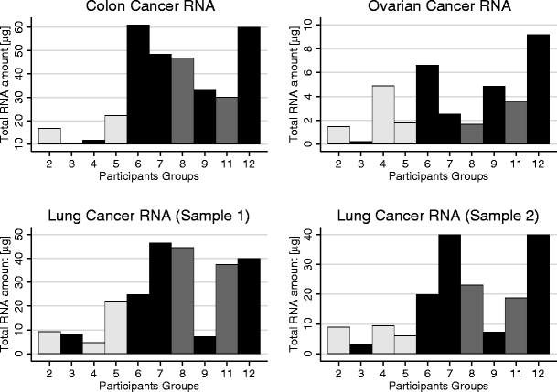
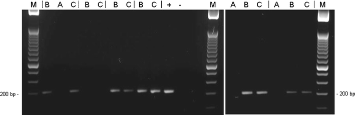
In most pathology laboratories worldwide, formalin-fixed paraffin embedded (FFPE) samples are the only tissue specimens available for routine diagnostics. Although commercial kits for diagnostic molecular pathology testing are becoming available, most of the current diagnostic tests are laboratory-based assays. Thus, there is a need for standardized procedures in molecular pathology, starting from the extraction of nucleic acids. To evaluate the current methods for extracting nucleic acids from FFPE tissues, 13 European laboratories, participating to the European FP6 program IMPACTS (www.impactsnetwork.eu), isolated nucleic acids from four diagnostic FFPE tissues using their routine methods, followed by quality assessment. The DNA-extraction protocols ranged from homemade protocols to commercial kits. Except for one homemade protocol, the majority gave comparable results in terms of the quality of the extracted DNA measured by the ability to amplify differently sized control gene fragments by PCR. For array-applications or tests that require an accurately determined DNA-input, we recommend using silica based adsorption columns for DNA recovery. For RNA extractions, the best results were obtained using chromatography column based commercial kits, which resulted in the highest quantity and best assayable RNA. Quality testing using RT-PCR gave successful amplification of 200 bp-250 bp PCR products from most tested tissues. Modifications of the proteinase-K digestion time led to better results, even when commercial kits were applied. The results of the study emphasize the need for quality control of the nucleic acid extracts with standardised methods to prevent false negative results and to allow data comparison among different diagnostic laboratories.


Figures

Fig. 1
Fig. 1
Bar graph representing the total amount of extracted DNA from colon cancer, ovarian cancer and lung cancer (2) specimens by the use of different protocol types. Light grey bars are referred to protocol type 1 (DNA extraction with precipitation step); medium grey bars are referred to protocol type 2 (DNA extraction without purifications steps) and dark grey bars are referred to protocol type 3 (DNA extraction with commercial kits based on the use of silica based columns)
Fig. 2
Fig. 2
BIOMED-2 control gene PCR results of the extracted DNAs in duplicate of group 9, 6 and 2 (protocol type 3, 2 and 1 respectively). DNA was extracted from ovary (a), lung 1 (b) and lung 2 (c). The size of the amplified products is indicated. The lowest thick band represents primer-dimer products
Fig. 3
Fig. 3
Bar graph representing the total amount of extracted RNA from colon cancer, ovarian cancer and lung cancer (2) specimens by the use of different protocol types. Light grey bars are referred to protocol type 4 (RNA extraction with home-made protocols); medium grey bars are referred to protocol type 5 (RNA extraction with monophasic commercial solutions) and dark grey bars are referred to protocol type 6 (RNA extraction with commercial kits based on the use of silica based columns)
Fig. 4
Fig. 4
B2M housekeeping gene specific RT-PCR results of the extracted RNAs of the group 2, 11, 6, 3, 7 and 12 (protocol type 4, 5 and four times 6, respectively). RNA was extracted from ovary (a), lung 1 (b) and lung 2 (c). The PCR products were separated in 3 % (w/v) agarose gels. The size of the amplification products is indicated (200 bp). M: size marker (50 bp DNA Ladder, Invitrogen, Karlsruhe, Germany)

References

    1. Dietel M. Predictive medicine: incipient reality or fata morgana? J Pathol. 2007;212:353–355. doi: 10.1002/path.2191. - DOI - PubMed
    1. Paik S, Tang G, Shak S, et al. Gene expression and benefit of chemotherapy in women with node-negative, estrogen receptor-positive breast cancer. J Clin Oncol. 2006;24:3726–3734. doi: 10.1200/JCO.2005.04.7985. - DOI - PubMed
    1. van Dongen JJ, Langerak AW, Bruggemann M, et al. Design and standardization of PCR primers and protocols for detection of clonal immunoglobulin and T-cell receptor gene recombinations in suspect lymphoproliferations: report of the BIOMED-2 Concerted Action BMH4-CT98-3936. Leukemia. 2003;17:2257–2317. doi: 10.1038/sj.leu.2403202. - DOI - PubMed
    1. Nardon E, Donada M, Bonin S, et al. Higher random oligo concentration improves reverse transcription yield of cDNA from bioptic tissues and quantitative RT-PCR reliability. Exp Mol Pathol. 2009;87:146–151. doi: 10.1016/j.yexmp.2009.07.005. - DOI - PubMed
    1. Masuda N, Ohnishi T, Kawamoto S, et al. Analysis of chemical modification of RNA from formalin-fixed samples and optimization of molecular biology applications for such samples. Nucleic Acids Res. 1999;27:4436–4443. doi: 10.1093/nar/27.22.4436. - DOI - PMC - PubMed

Publication types