. 2010 Nov 15;58(15):1827-37.
doi: 10.1002/glia.21052.

Injury and differentiation following inhibition of mitochondrial respiratory chain complex IV in rat oligodendrocytes

Affiliations
Free PMC article

Injury and differentiation following inhibition of mitochondrial respiratory chain complex IV in rat oligodendrocytes

Iryna Ziabreva et al. Glia. .
Free PMC article

Abstract

Oligodendrocyte lineage cells are susceptible to a variety of insults including hypoxia, excitotoxicity, and reactive oxygen species. Demyelination is a well-recognized feature of several CNS disorders including multiple sclerosis, white matter strokes, progressive multifocal leukoencephalopathy, and disorders due to mitochondrial DNA mutations. Although mitochondria have been implicated in the demise of oligodendrocyte lineage cells, the consequences of mitochondrial respiratory chain defects have not been examined. We determine the in vitro impact of established inhibitors of mitochondrial respiratory chain complex IV or cytochrome c oxidase on oligodendrocyte progenitor cells (OPCs) and mature oligodendrocytes as well as on differentiation capacity of OPCs from P0 rat. Injury to mature oligodendrocytes following complex IV inhibition was significantly greater than to OPCs, judged by cell detachment and mitochondrial membrane potential (MMP) changes, although viability of cells that remained attached was not compromised. Active mitochondria were abundant in processes of differentiated oligodendrocytes and MMP was significantly greater in differentiated oligodendrocytes than OPCs. MMP dissipated following complex IV inhibition in oligodendrocytes. Furthermore, complex IV inhibition impaired process formation within oligodendrocyte lineage cells. Injury to and impaired process formation of oligodendrocytes following complex IV inhibition has potentially important implications for the pathogenesis and repair of CNS myelin disorders.


Figures

Figure 1
Figure 1
Experimental timeline. To investigate injury to and detachment of cells following complex IV inhibition, P0 rat OPCs were proliferated for 2 days with PDGF and FGF and then differentiated for 5 days prior to exposure to sodium azide (1, 10, and 100 μM) or potassium cyanide (10 μM and 100 μM). The differentiation capacity of OPCs was assessed by either persistent or brief exposure to complex IV inhibitor at concentrations that did not reduce the number of OPCs (up to 10 μM). In persistent exposure experiments, complex IV inhibitor was added at the onset of OPC differentiation and kept throughout differentiation (persistent exposure) in the media, 25% of which was replaced on Day 4. To rule out the possibility that oligodendrocytes differentiate and then lost rather than fail to differentiate in persistent exposure experiments, OPCs were briefly exposed to sodium azide for 10 min and then washed prior to differentiation (brief exposure). The control samples, appropriate for the time points analyzed, were not exposed to the inhibitor. I: complex IV inhibitor (sodium azide or potassium cyanide).
Figure 2
Figure 2
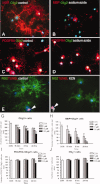
Injury to oligodendrocyte lineage cells from P0 rat following complex IV inhibition. A, C, and E: There were mature oligodendrocytes expressing MBP (a) and OPCs expressing PDGFRA (c) or NG2 (e) in oligodendrocyte cultures, derived from purified OPCs proliferated for 2 days and differentiated for 5 days. B, D, and F: Injury to processes of oligodendrocyte lineage cells and loss of mature oligodendrocytes (b) were the most striking changes observed following inhibition of complex IV. Images show changes following 100 μM of sodium azide for 3 h (b and d) and 100 μM of potassium cyanide for 3 h. TUNEL positive nuclei (e and f, arrowheads) were detected on average in 13.7% of oligodendrocyte lineage cells, which did not change significantly following complex IV inhibition. GJ: The quantitation of oligodendrocyte lineage cells remaining attached in the chamber slides showed a concentration dependent reduction in absolute number of Olig2+ nuclei (g). Loss of Olig2+ cells was confirmed by DAPI staining. Mature oligodendrocytes were injured and detached following complex IV inhibition in a dose dependent manner (h). In contrast, OPCs (PDGFRA+ or NG2+) remained attached to the chamber slides and the numbers were not effected by complex IV inhibition, except with 100 μM sodium azide at 24 h (i and j). The experiments, controls, and different concentrations of inhibitors in triplicate chambers, were performed on 10 separate occasions from six different litters. *P < 0.001. #P < 0.01. [Color figure can be viewed in the online issue, which is available at wileyonlinelibrary.com.]
Figure 3
Figure 3
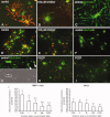
Detached oligodendrocytes in supernatant reflect cell loss following complex IV inhibition and express caspase 9 and translocate AIF to nucleus. A: The density of oligodendrocyte lineage cells in the supernatant, later captured on cytospins, increases in a dose dependent manner following exposure to sodium azide, reflecting the extent of cell loss in chamber slides (Fig. .2e). A density of 10 cells per μL in 400 μL of supernatant from one chamber represents detachment of 32% of cells. B and C: Detached MBP expressing cells captured on cytospins express caspase 9 (b) and show evidence of nuclear translocation of AIF (c, arrowheads). AIF positive cells without MBP expression (arrow) are most likely detached OPCs. Co-staining of caspase 9 and AIF was not possible as both primary antibodies were raised in rabbit. D: The extent of caspase 9 expression in detached cells varies in a dose and time dependent manner, with the majority of MBP expressing cells expressing caspase 9 when exposed to 100 μM of sodium azide. Caspase 9 was not detected in approximately half of the detached MBP expressing cells in control chambers or following exposure to 1 μM of sodium azide. The experiments, controls, and different concentrations of inhibitors in triplicates, were performed on four occasions from different litters. *P < 0.001. #P < 0.01. [Color figure can be viewed in the online issue, which is available at wileyonlinelibrary.com.]
Figure 4
Figure 4
Mitochondrial membrane potential in live oligodendrocyte lineage cells from P0 rat. AI: Mitochondrial membrane potential (MMP) was determined using JC-1. Oligodendrocyte processes in cultures differentiated for 5 days (a) contained strikingly more active mitochondria (red) than processes of OPCs proliferated without differentiation for 5 days (d). Exposure of differentiated oligodendrocytes and OPCs to complex IV inhibitor (shown following 10 μM of sodium azide exposure for 15 min) led to a notable dissipation of MMP (b and e). Non-aggregated JC-1 is shown by green fluorescence (a–b and d–e). The abundance of mitochondria, judged by JC-1 aggregation, in the processes of differentiated oligodendrocytes (c) relative to undifferentiated OPCs (f) was confirmed using MitoTraker green. The tips of differentiating oligodendrocytes, with bulbous morphology on brightfield image (g), were particularly notable for the presence of mitochondria (g, arrowheads). The mitochondrial membrane uncoupler, carbonylcyanide-p-trifluoromethoxyphenylhydrazone (FCCP), dissipated MMP in differentiated oligodendrocytes (h), and OPCs (i) as expected. J: Quantitation of MMP based on ratio of red to green fluorescence intensity showed the MMP to be much greater in differentiated oligodendrocytes compared with OPCs in controls. MMP decreased significantly in differentiated oligodendrocytes when exposed to 1 μM of sodium azide for 15 min whereas MMP in OPCs was less susceptible to low doses (1 μM and 10 μM) of complex IV inhibitor. *P = 0.001. **P < 0.001. #P = 0.023. [Color figure can be viewed in the online issue, which is available at wileyonlinelibrary.com.]
Figure 5
Figure 5
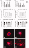
Density and morphology of mature oligodendrocytes when P0 rat OPCs were persistently or briefly exposed to complex IV inhibitor during or prior to differentiation. AC: When proliferated OPCs were persistently exposed to sodium azide throughout the 5-day-period of differentiation (Fig. .1) at concentrations that did not effect the viability (up to 10 μM of sodium azide), the total number of oligodendrocyte lineage cells (MBP+, PDGFRA+, or NG2+) remaining in the chamber slides is similar compared with controls (a). With 10 μM of sodium azide there was a significant reduction in the percentage of MBP+ cells reflecting either impaired differentiation or injury to differentiated MBP+ cells (b). When mature oligodendrocytes were categorized into rounded (category I, GI), simple processes (category II, GII), complex processes (category III, GIII), or with MBP+ membrane (category IV, GIV) the proportion of MBP+ cells lacking processes or with simple processes was significantly greater when differentiated in the presence of complex IV inhibitor compared with controls (c). DF: Brief exposure of OPCs to variable concentrations of sodium azide prior to differentiation (Fig. .1) led to similar number of oligodendrocyte lineage cells compared with controls (d). We did not detect a significant change in the number of MBP+ cells in brief exposure experiments (e). There was a significant increase in MBP expressing cells without processes or with simple processes in brief exposure experiments (f). The experiments, controls, and different concentrations of inhibitors in triplicate chambers, were performed on eight occasions from different litters. *P < 0.01. **P < 0.001. [Color figure can be viewed in the online issue, which is available at wileyonlinelibrary.com.]

References

    1. Aboul-Enein F, Rauschka H, Kornek B, Stadelmann C, Stefferl A, Bruck W, Lucchinetti C, Schmidbauer M, Jellinger K, Lassmann H. Preferential loss of myelin-associated glycoprotein reflects hypoxia-like white matter damage in stroke and inflammatory brain diseases. J Neuropathol Exp Neurol. 2003;62:25–33. - PubMed
    1. Adam-Vizi V. Production of reactive oxygen species in brain mitochondria: Contribution by electron transport chain and non-electron transport chain sources. Antioxid Redox Signal. 2005;7:1140–1149. - PubMed
    1. Almazan G, Liu HN, Khorchid A, Sundararajan S, Martinez-Bermudez AK, Chemtob S. Exposure of developing oligodendrocytes to cadmium causes HSP72 induction, free radical generation, reduction in glutathione levels, and cell death. Free Radic Biol Med. 2000;29:858–869. - PubMed
    1. Andrews T, Zhang P, Bhat NR. TNFalpha potentiates IFNgamma-induced cell death in oligodendrocyte progenitors. J Neurosci Res. 1998;54:574–583. - PubMed
    1. Back SA, Gan X, Li Y, Rosenberg PA, Volpe JJ. Maturation-dependent vulnerability of oligodendrocytes to oxidative stress-induced death caused by glutathione depletion. J Neurosci. 1998;18:6241–6253. - PMC - PubMed

Publication types

MeSH terms