

Crystal structure of Bacillus subtilis SPP1 phage gp23.1, a putative chaperone
- PMID: 20665904
- PMCID: PMC2975145
- DOI: 10.1002/pro.464
Crystal structure of Bacillus subtilis SPP1 phage gp23.1, a putative chaperone
Abstract
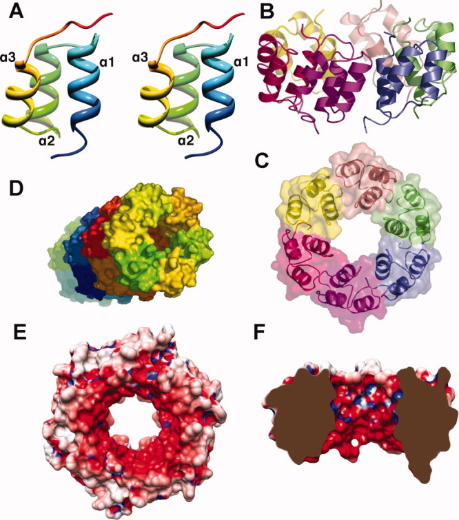
SPP1 is a siphophage infecting the gram-positive bacterium Bacillus subtilis. The SPP1 tail electron microscopy (EM) reconstruction revealed that it is mainly constituted by conserved structural proteins such as the major tail proteins (gp17.1), the tape measure protein (gp18), the Distal tail protein (Dit, gp19.1), and the Tail associated lysin (gp21). A group of five small genes (22-24.1) follows in the genome but it remains to be elucidated whether their protein products belong or not to the tail. Noteworthy, an unassigned EM density accounting for ~245 kDa is present at the distal end of the SPP1 tail-tip. We report here the gp23.1 crystal structure at 1.6 A resolution, a protein that lacks sequence identity to any known protein. We found that gp23.1 forms a hexamer both in the crystal lattice and in solution as revealed by light scattering measurements. The gp23.1 hexamer does not fit well in the unassigned SPP1 tail-tip EM density and we hypothesize that this protein might act as a chaperone.
Copyright © 2010 The Protein Society.
Figures
References
-
- Brussow H, Hendrix RW. Phage genomics: small is beautiful. Cell. 2002;108:13–16. - PubMed
-
- Tavares P, Santos MA, Lurz R, Morelli G, de Lencastre H, Trautner TA. Identification of a gene in Bacillus subtilis bacteriophage SPP1 determining the amount of packaged DNA. J Mol Biol. 1992;225:81–92. - PubMed
-
- Sao-Jose C, Lhuillier S, Lurz R, Melki R, Lepault J, Santos MA, Tavares P. The ectodomain of the viral receptor YueB forms a fiber that triggers ejection of bacteriophage SPP1 DNA. J Biol Chem. 2006;281:11464–11470. - PubMed
Publication types
MeSH terms
Substances
Grants and funding
LinkOut - more resources
Full Text Sources
Molecular Biology Databases
Research Materials
Miscellaneous
