

Relationship between knee and ankle degeneration in a population of organ donors
- PMID: 20667091
- PMCID: PMC2925325
- DOI: 10.1186/1741-7015-8-48
Relationship between knee and ankle degeneration in a population of organ donors
Abstract
Background: Osteoarthritis (OA) is a progressive degenerative condition of synovial joints in response to both internal and external factors. The relationship of OA in one joint of an extremity to another joint within the same extremity, or between extremities, has been a topic of interest in reference to the etiology and/or progression of the disease.
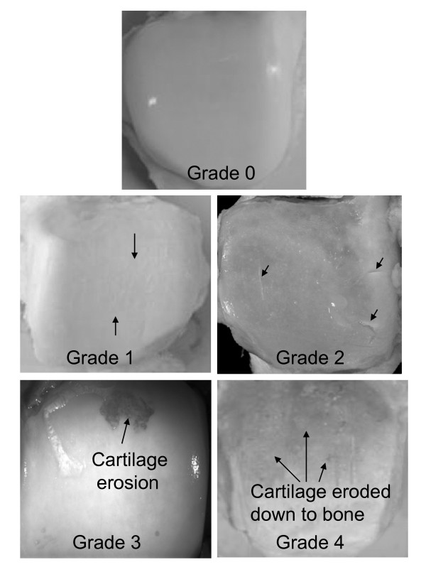
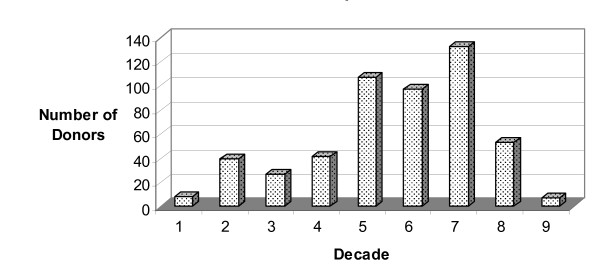
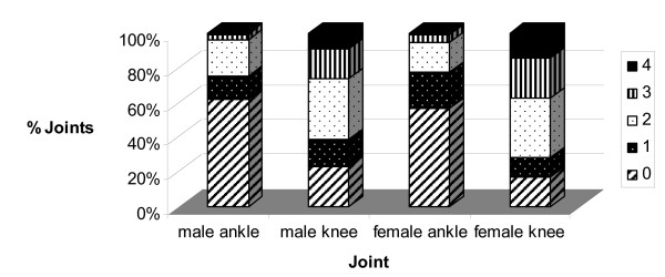
Methods: The prevalence of articular cartilage lesions and osteophytes, characteristic of OA, was evaluated through visual inspection and grading in 1060 adult knee/tali pairs from 545 cadaveric joint donors.
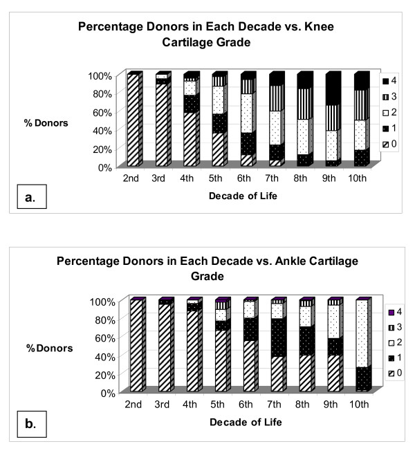
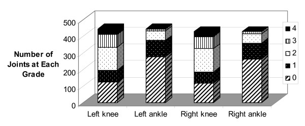
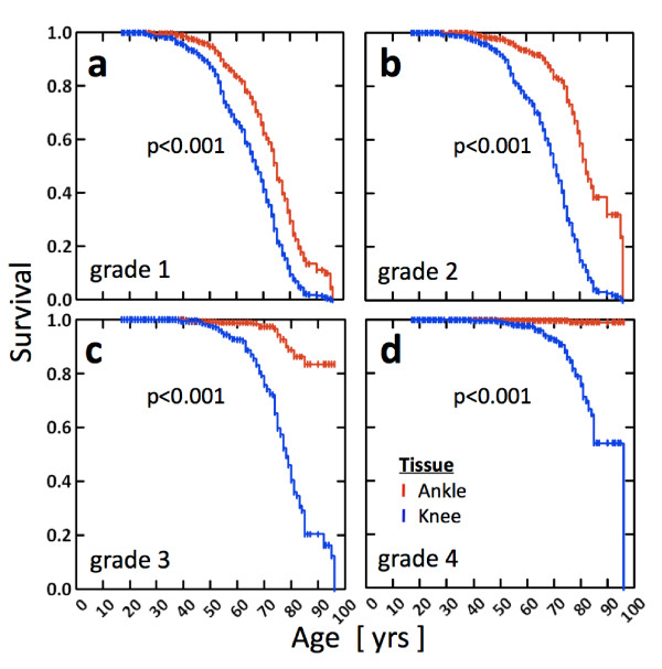
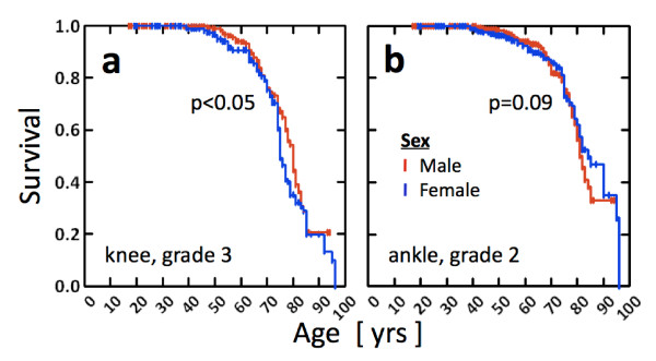
Results: Joint degeneration increased more rapidly with age for the knee joint, and significantly more knee joints displayed more severe degeneration than ankle joints from as early as the third decade. Women displayed more severe knee degeneration than did men. Severe ankle degeneration did not exist in the absence of severe knee degeneration. The effect of weight on joint degeneration was joint-specific whereby weight had a significantly greater effect on the knee. Ankle grades increasingly did not match within a donor as the grade of degeneration in either the left or the right knee increased.
Conclusions: Gender and body type have a greater effect on knee joint integrity as compared to the ankle, suggesting that knees are more prone to internal causative effects of degeneration. We hypothesize that the greater variability in joint health between joints within an individual as disease progresses from normal to early signs of degeneration may be a result of mismatched limb kinetics, which in turn might lead to joint disease progression.
Figures

Similar articles
-
Prevalence of articular cartilage degeneration in the ankle and knee joints of human organ donors.J Orthop Sci. 1999;4(6):407-12. doi: 10.1007/s007760050123. J Orthop Sci. 1999. PMID: 10664423
-
Distinguishing ankle and knee articular cartilage.Foot Ankle Clin. 2003 Jun;8(2):305-16, x. doi: 10.1016/s1083-7515(03)00012-3. Foot Ankle Clin. 2003. PMID: 12911243
-
Cartilage calcification of the ankle joint is associated with osteoarthritis in the general population.BMC Musculoskelet Disord. 2018 May 24;19(1):169. doi: 10.1186/s12891-018-2094-7. BMC Musculoskelet Disord. 2018. PMID: 29793463 Free PMC article.
-
Osteoarthritis in ankle and knee joints.Semin Arthritis Rheum. 1997 Feb;26(4):667-74. doi: 10.1016/s0049-0172(97)80002-9. Semin Arthritis Rheum. 1997. PMID: 9062947 Review.
-
Cartilage degeneration in different human joints.Osteoarthritis Cartilage. 2005 Feb;13(2):93-103. doi: 10.1016/j.joca.2004.11.006. Osteoarthritis Cartilage. 2005. PMID: 15694570 Review.
Cited by
-
Atlas of radiographic features of osteoarthritis of the ankle and hindfoot.Osteoarthritis Cartilage. 2015 Dec;23(12):2059-2085. doi: 10.1016/j.joca.2015.08.008. Epub 2015 Aug 28. Osteoarthritis Cartilage. 2015. PMID: 26318654 Free PMC article. Review.
-
Association between the Body Roundness Index and osteoarthritis: A population-based study.PLoS One. 2025 Jun 18;20(6):e0326334. doi: 10.1371/journal.pone.0326334. eCollection 2025. PLoS One. 2025. PMID: 40531865 Free PMC article.
-
Local and systemic cardiovascular effects from monochromatic infrared therapy in patients with knee osteoarthritis: a double-blind, randomized, placebo-controlled study.Evid Based Complement Alternat Med. 2012;2012:583016. doi: 10.1155/2012/583016. Epub 2012 Jun 27. Evid Based Complement Alternat Med. 2012. PMID: 22792125 Free PMC article.
-
The epidemiology of sleep and obesity.Sleep Health. 2017 Oct;3(5):383-388. doi: 10.1016/j.sleh.2017.07.013. Epub 2017 Aug 15. Sleep Health. 2017. PMID: 28923198 Free PMC article. Review.
-
Ankle alignment before and after total knee arthroplasty in patients with valgus knee deformity.J Orthop Surg Res. 2025 Apr 18;20(1):389. doi: 10.1186/s13018-025-05800-5. J Orthop Surg Res. 2025. PMID: 40247287 Free PMC article.
References
-
- Issa SN, Dunlop D, Chang A, Song J, Prasad PV, Guermazi A, Peterfy C, Cahue S, Marshall M, Kapoor D, Hayes K, Sharma L. Full-limb and knee radiography assessments of varus-valgus alignment and their relationship to osteoarthritis disease features by magnetic resonance imaging. Arthritis Rheum. 2008;57:398–406. doi: 10.1002/art.22618. - DOI - PubMed
