. 2010 Jul 28:10:230.
doi: 10.1186/1471-2148-10-230.

Interference with histidyl-tRNA synthetase by a CRISPR spacer sequence as a factor in the evolution of Pelobacter carbinolicus

Affiliations

Interference with histidyl-tRNA synthetase by a CRISPR spacer sequence as a factor in the evolution of Pelobacter carbinolicus

Muktak Aklujkar et al. BMC Evol Biol. .

Abstract

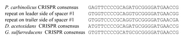
Background: Pelobacter carbinolicus, a bacterium of the family Geobacteraceae, cannot reduce Fe(III) directly or produce electricity like its relatives. How P. carbinolicus evolved is an intriguing problem. The genome of P. carbinolicus contains clustered regularly interspaced short palindromic repeats (CRISPR) separated by unique spacer sequences, which recent studies have shown to produce RNA molecules that interfere with genes containing identical sequences.

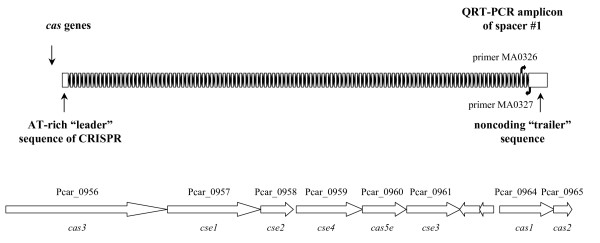
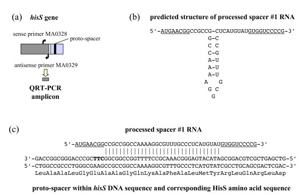
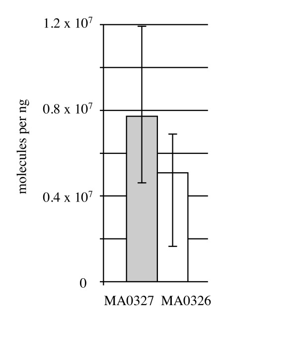
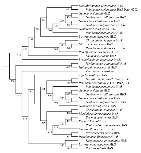
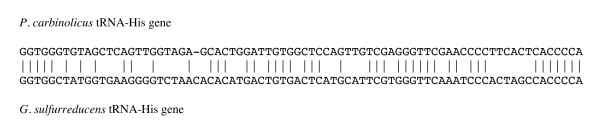
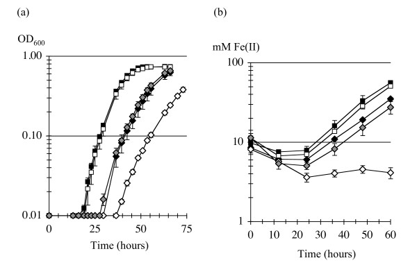
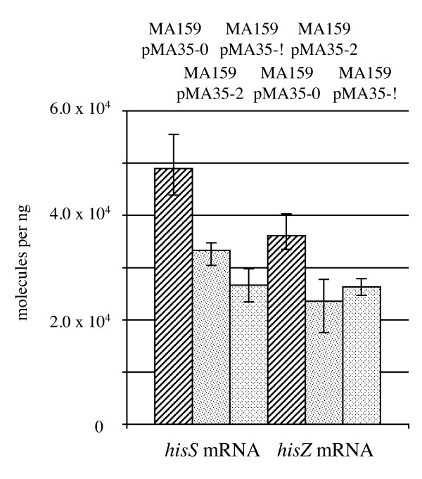
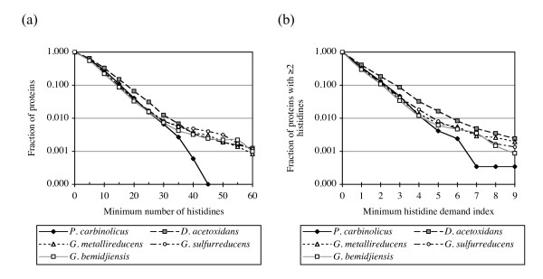
Results: CRISPR spacer #1, which matches a sequence within hisS, the histidyl-tRNA synthetase gene of P. carbinolicus, was shown to be expressed. Phylogenetic analysis and genetics demonstrated that a gene paralogous to hisS in the genomes of Geobacteraceae is unlikely to compensate for interference with hisS. Spacer #1 inhibited growth of a transgenic strain of Geobacter sulfurreducens in which the native hisS was replaced with that of P. carbinolicus. The prediction that interference with hisS would result in an attenuated histidyl-tRNA pool insufficient for translation of proteins with multiple closely spaced histidines, predisposing them to mutation and elimination during evolution, was investigated by comparative genomics of P. carbinolicus and related species. Several ancestral genes with high histidine demand have been lost or modified in the P. carbinolicus lineage, providing an explanation for its physiological differences from other Geobacteraceae.

Conclusions: The disappearance of multiheme c-type cytochromes and other genes typical of a metal-respiring ancestor from the P. carbinolicus lineage may be the consequence of spacer #1 interfering with hisS, a condition that can be reproduced in a heterologous host. This is the first successful co-introduction of an active CRISPR spacer and its target in the same cell, the first application of a chimeric CRISPR construct consisting of a spacer from one species in the context of repeats of another species, and the first report of a potential impact of CRISPR on genome-scale evolution by interference with an essential gene.


Figures

Figure 1
Figure 1
The CRISPR locus of P. carbinolicus. This locus consists of 112 repeats (black diamonds) separated by 111 nonrepetitive spacers (white rectangles). Spacer #1 is at the trailer end, farthest from the cas genes and the AT-rich leader sequence near which new spacers are typically inserted. Primers MA0326 and MA0327 are based on sequences surrounding spacer #1, and were used to detect its RNA transcript. The arrangement of the cas genes (located immediately to the left of the leader sequence) is illustrated in the lower half of the figure. The two intervening genes encode a putative toxin (Pcar_0962) and transcriptional regulator or antitoxin (Pcar_0963).
Figure 2
Figure 2
CRISPR spacer #1 matches a nucleotide sequence within the hisS gene. (a) hisS consists of a catalytic domain (dark grey) and an anticodon loop recognition domain (light grey) connected by a linker (white stripe). The proto-spacer sequence matching spacer #1 (black stripe) is within the anticodon loop recognition domain. Primers MA0328 and MA0329 were designed to amplify a cDNA segment from the catalytic domain. (b) Predicted secondary structure of a processed CRISPR transcript (initiated at the leader sequence) that contains spacer #1, before hybridization to hisS DNA. Sequences from the repeats flanking spacer #1 are underlined. (c) Predicted hybridization of a proto-spacer segment within the anticodon loop recognition domain of hisS DNA (template strand) with a processed spacer #1 RNA. The proto-spacer-adjacent motif CTT is shown in bold.
Figure 3
Figure 3
Spacer #1 is transcribed into RNA in P. carbinolicus, with both strands similarly abundant. Reverse transcription was performed with either primer MA0327 (grey bar) or primer MA0326 (white bar), and the amount of cDNA was quantified by QRT-PCR. The mean of three biological replicates is shown; error bars represent the minimum and maximum.
Figure 4
Figure 4
Phylogeny of HisS and HisZ proteins. Pcar_1041 and orthologous proteins of Geobacteraceae cluster among true histidyl-tRNA synthetases (HisS), whereas Pcar_0202 and its orthologs cluster among the HisZ proteins, which are the regulatory subunit of ATP phosphoribosyltransferase. Confidence values are out of 500 bootstraps.
Figure 5
Figure 5
Alignment of the sequences of tRNA-His genes from P. carbinolicus and G. sulfurreducens.
Figure 6
Figure 6
Growth of G. sulfurreducens with hisS of P. carbinolicus is inhibited by spacer #1. (a) Growth on NBAF medium by reduction of fumarate. (b) Growth on FWAFC medium by reduction of Fe(III) citrate. The strains shown are wild type G. sulfurreducens DL1 (black squares); DL1(pMA35-2) with two copies of spacer #1 in a plasmid-borne chimeric CRISPR (white squares); transgenic strain MA159, which has hisS of P. carbinolicus (black diamonds); MA159(pMA35-2) with both the hisS transgene and two copies of spacer #1 (white diamonds); and MA159(pMA35-0) with the hisS transgene and a CRISPR repeat without spacer #1 (grey diamonds).
Figure 7
Figure 7
Spacer #1 has similar effects on the amounts of hisS and hisZ RNA. The strains shown are G. sulfurreducens MA159(pMA35-0) with the hisS transgene and a CRISPR repeat without spacer #1 (diagonally striped bars); MA159(pMA35-2) with both the hisS transgene and two copies of spacer #1 (speckled bars); and MA159(pMA35-!) with the hisS transgene and a single mutated copy of spacer #1 (diamond-patterned bars). Reverse transcription was performed with either primer MA0329 for hisS or primer MA0442 for hisZ, and the amount of cDNA was quantified by QRT-PCR. The mean of three biological replicates is shown; error bars represent the minimum and maximum.
Figure 8
Figure 8
The P. carbinolicus genome encodes fewer proteins with multiple closely spaced histidines. (a) The fraction of proteins with a given minimum number of histidines, plotted for the genomes of P. carbinolicus (black diamonds), D. acetoxidans (grey squares), G. sulfurreducens (white circles), G. metallireducens (white triangles) and G. bemidjiensis (white squares). (b) The fraction of proteins with two or more histidines and a given minimum histidine demand index, plotted for the same five genomes.
Figure 9
Figure 9
Alignment of the repeats on either side of spacer #1 with the CRISPR consensus sequences of P. carbinolicus, D. acetoxidans and G. sulfurreducens.

Similar articles

Cited by

References

    1. Godde JS, Bickerton A. The repetitive DNA elements called CRISPRs and their associated genes: evidence of horizontal transfer among prokaryotes. J Mol Evol. 2006;62(6):718–729. doi: 10.1007/s00239-005-0223-z. - DOI - PubMed
    1. Grissa I, Vergnaud G, Pourcel C. The CRISPRdb database and tools to display CRISPRs and to generate dictionaries of spacers and repeats. BMC Bioinformatics. 2007;8:172. doi: 10.1186/1471-2105-8-172. - DOI - PMC - PubMed
    1. Haft DH, Selengut J, Mongodin EF, Nelson KE. A guild of 45 CRISPR-associated (Cas) protein families and multiple CRISPR/Cas subtypes exist in prokaryotic genomes. PLoS Comput Biol. 2005;1(6):e60. doi: 10.1371/journal.pcbi.0010060. - DOI - PMC - PubMed
    1. Jansen R, Embden JD, Gaastra W, Schouls LM. Identification of genes that are associated with DNA repeats in prokaryotes. Mol Microbiol. 2002;43(6):1565–1575. doi: 10.1046/j.1365-2958.2002.02839.x. - DOI - PubMed
    1. Lillestøl RK, Redder P, Garrett RA, Brugger K. A putative viral defence mechanism in archaeal cells. Archaea. 2006;2(1):59–72. doi: 10.1155/2006/542818. - DOI - PMC - PubMed

Publication types

LinkOut - more resources