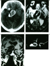
Giant serpentine aneurysms: multidisciplinary management. Report of four cases and review of the literature
- PMID: 20667180
- PMCID: PMC3679576
- DOI: 10.1177/159101990000600105
Giant serpentine aneurysms: multidisciplinary management. Report of four cases and review of the literature
Abstract
Sixty-five cases of intracranial giant serpentine aneurysms (GSAs), including 61 cases reported in the literature and four additional cases presented in this study were reviewed. The clinical presentation, possible causes, natural history, and especially management of GSAs are discussed with emphasis on the need for aggressive intervention and multidisciplinary management.
Figures


Similar articles
-
Flow diversion treatment for giant intracranial serpentine aneurysms.Front Aging Neurosci. 2022 Nov 3;14:988411. doi: 10.3389/fnagi.2022.988411. eCollection 2022. Front Aging Neurosci. 2022. PMID: 36408107 Free PMC article.
-
Endovascular occlusion of giant serpentine aneurysm: A case report and literature review.J Cerebrovasc Endovasc Neurosurg. 2022 Mar;24(1):51-57. doi: 10.7461/jcen.2022.E2021.06.003. Epub 2022 Jan 14. J Cerebrovasc Endovasc Neurosurg. 2022. PMID: 35026888 Free PMC article.
-
Cerebral giant serpentine aneurysm: case report and review of the literature.Neurosurgery. 1988 Jul;23(1):92-7. doi: 10.1227/00006123-198807000-00016. Neurosurgery. 1988. PMID: 3050586 Review.
-
Endovascular trapping of giant serpentine aneurysms by using Guglielmi detachable coils: successful reduction of mass effect. Report of two cases.J Neurosurg. 2001 May;94(5):836-40. doi: 10.3171/jns.2001.94.5.0836. J Neurosurg. 2001. PMID: 11354420
-
Giant serpentine aneurysms: a review and presentation of five cases.AJNR Am J Neuroradiol. 1995 May;16(5):1061-72. AJNR Am J Neuroradiol. 1995. PMID: 7639128 Free PMC article. Review.
Cited by
-
A giant partial thrombosed aneurysm of the internal cavernous carotid artery mimicking a meningioma of the lesser wing of the sphenoid bone.Radiol Case Rep. 2022 Feb 19;17(4):1325-1329. doi: 10.1016/j.radcr.2022.01.075. eCollection 2022 Apr. Radiol Case Rep. 2022. PMID: 35242260 Free PMC article.
-
Comprehensive Evaluation of Serpentine Aneurysms: a Systematic Review and Meta-analysis with a Subanalysis for Treatment Approaches.Clin Neuroradiol. 2024 Dec;34(4):749-760. doi: 10.1007/s00062-024-01460-w. Epub 2024 Sep 24. Clin Neuroradiol. 2024. PMID: 39316117 English.
-
Giant serpentine internal carotid artery aneurysm: endovascular parent artery occlusion. A pediatric case report.Interv Neuroradiol. 2007 Mar;13(1):85-94. doi: 10.1177/159101990701300112. Epub 2007 Jun 27. Interv Neuroradiol. 2007. PMID: 20566135 Free PMC article.
-
A comprehensive analysis and systematic review of giant serpentine aneurysms.Ann Med Surg (Lond). 2025 Feb 26;87(3):1458-1466. doi: 10.1097/MS9.0000000000003000. eCollection 2025 Mar. Ann Med Surg (Lond). 2025. PMID: 40213197 Free PMC article. Review.
-
Study and Therapeutic Progress on Intracranial Serpentine Aneurysms.Int J Med Sci. 2016 May 26;13(6):432-9. doi: 10.7150/ijms.14934. eCollection 2016. Int J Med Sci. 2016. PMID: 27279792 Free PMC article. Review.
References
-
- Segal HD, Mclaurin RL. Giant serpentine aneurysm. Report of two cases. J Neurosurg. 1977;46:115–120. - PubMed
-
- Suzuki S, Takahashi T, et al. Management of giant serpentine aneurysms of the middle cerebral artery-review of literature and report of a case successfully treated by STA-MCA anastomosis only. Acta Neurochir (Wien) 1992;117:23–29. - PubMed
-
- Patel DV, Sherman IC, et al. Giant serpentine intracranial aneurysm. Surg Neurol. 1981;16:402–407. - PubMed
-
- Haddad GF, Haddad FS. Cerebral giant serpentine aneurysm: case report and review of the literature. Neurosurgery. 1988;23:92–96. - PubMed
LinkOut - more resources
Full Text Sources
Miscellaneous

