

Palliative embolisation of brain arteriovenous malformations presenting with progressive neurological deficit
- PMID: 20667196
- PMCID: PMC3679675
- DOI: 10.1177/159101990000600302
Palliative embolisation of brain arteriovenous malformations presenting with progressive neurological deficit
Abstract
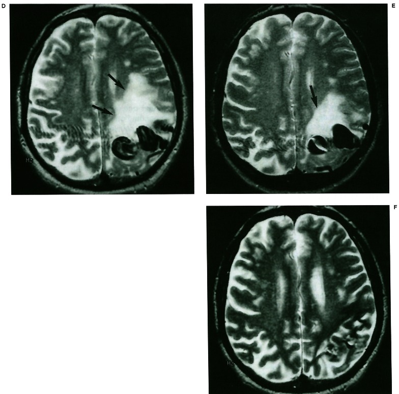
Large arteriovenous malformations (AVMs) located in eloquent areas of the brain are generally considered incurable because of the high morbidity and mortality associated with their treatment. When these patients develop a progressive neurological deficit they in time often become severely disabled. This report presents the results of palliative embolisation in this subgroup of patients. Analysis of our data-base of 714 patients with known brain AVMs revealed 17 patients who presented with progressive neurological deficit and who underwent palliative embolisation as the therapeutic modality of choice for management of their AVM. One patient was excluded due to lack of follow-up and two were excluded because they later received radiation therapy. Following embolisation 43% had improvement of their neurological deficit, 50% stabilized and 7% continued to deteriorate and these clinical results persisted for an average of more than 2 years follow-up. Transient neurological morbidity associated with embolisation treatment was 7% and there was no permanent morbidity and no mortality. Palliative embolisation of brain AVMs presenting with progressive neurological deficits arrested deterioration in more than 90% of patients and was associated with low morbidity and no mortality.
Figures


Similar articles
-
Microsurgery for cerebral arteriovenous malformations: subgroup outcomes in a consecutive series of 288 cases.J Neurosurg. 2017 Apr;126(4):1056-1063. doi: 10.3171/2016.4.JNS153017. Epub 2016 Jun 10. J Neurosurg. 2017. PMID: 27285541 Clinical Trial.
-
Management of patients with brain arteriovenous malformations.Eur J Radiol. 2003 Jun;46(3):195-205. doi: 10.1016/s0720-048x(03)00091-3. Eur J Radiol. 2003. PMID: 12758114 Review.
-
Ventricular/paraventricular small arteriovenous malformations: role of embolisation with cyanoacrylate.Neuroradiology. 2005 Apr;47(4):287-94. doi: 10.1007/s00234-005-1339-y. Epub 2005 Apr 2. Neuroradiology. 2005. PMID: 15806431
-
Changing role for preoperative embolisation in the management of arteriovenous malformations of the brain.J Clin Neurosci. 2000 Nov;7(6):527-30. doi: 10.1054/jocn.2000.0759. J Clin Neurosci. 2000. PMID: 11029234
-
Microsurgical treatment of arteriovenous malformations: analysis and comparison with stereotactic radiosurgery.J Neurosurg. 1998 Apr;88(4):641-6. doi: 10.3171/jns.1998.88.4.0641. J Neurosurg. 1998. PMID: 9525708 Review.
Cited by
-
Endovascular treatment of B-AVM.Interv Neuroradiol. 2003 Oct 10;9(Suppl 2):109-11. doi: 10.1177/15910199030090S221. Epub 2004 Oct 22. Interv Neuroradiol. 2003. PMID: 20591294 Free PMC article. No abstract available.
-
Brain edema associated with unruptured brain arteriovenous malformations.Neuroradiology. 2009 May;51(5):327-35. doi: 10.1007/s00234-009-0500-4. Epub 2009 Feb 15. Neuroradiology. 2009. PMID: 19219601
References
-
- Richling B, Bavinzski G. Arterio-venous malformations of the basal ganglia. Surgical versus endovascular treatment. Acta Neurochir Supple. 1991;53:50–59. - PubMed
-
- Tew JM, Lewis AI, Reichert KW. Management strategies and surgical techniques for deep-seated supratentorial arteriovenous malformations. Neurosurgery. 1995;36:1065–1072. - PubMed
-
- Yamada S. Surgical approach to arteriovenous malformations in functional areas of the brain. In: Yamada S, editor. Arteriovenous malformations in functional areas of the brain. Armonk, NY: Futura; 1999. pp. 1–123. In: Yamada S, (ed)
-
- Brown RD, Wiebers DO, et al. The natural history of unruptured intracranial arteriovenous malformations. J Neurosurg. 1988;68:352–357. - PubMed
-
- Ondra SL, Troupp H, et al. The natural history of symptomatic arteriovenous malformation of the brain: a 24-year follow-up assessment. J Neurosurg. 1990;73:387–391. - PubMed
LinkOut - more resources
Full Text Sources
