The arteriovenous malformation associated with major arterial occlusion and moyamoya vessels: a cerebral blood flow study
- PMID: 20667197
- PMCID: PMC3679676
- DOI: 10.1177/159101990000600303
The arteriovenous malformation associated with major arterial occlusion and moyamoya vessels: a cerebral blood flow study
Abstract
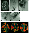
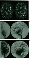
We report 2 patients with arteriovenous malformation (AVM) associated with complete occlusion of the unilateral middle cerebral artery and moyamoya vessels. Xenon CT CBF study demonstrated diffusely decreased CMF in unilateral or bilateral hemispheres with multiple areas of decreased vascular reserve. A significant reduction of AVM size was seen in one patient who received radiosurgery with marked CBF improvement.
Figures


Similar articles
-
Arteriovenous malformation with an occlusive feeding artery coexisting with unilateral moyamoya disease.J Clin Neurol. 2010 Dec;6(4):216-20. doi: 10.3988/jcn.2010.6.4.216. Epub 2010 Dec 31. J Clin Neurol. 2010. PMID: 21264203 Free PMC article.
-
Use of acetazolamide-challenge xenon CT in the assessment of cerebral blood flow dynamics in patients with arteriovenous malformations.AJNR Am J Neuroradiol. 1990 May;11(3):441-8. AJNR Am J Neuroradiol. 1990. PMID: 2112305 Free PMC article.
-
Association of cerebral arteriovenous malformations and spontaneous occlusion of major feeding arteries: clinical and therapeutic implications.Neurosurgery. 1999 Nov;45(5):1105-11; discussion 1111-2. doi: 10.1097/00006123-199911000-00018. Neurosurgery. 1999. PMID: 10549926 Review.
-
Development of a de novo arteriovenous malformation after bilateral revascularization surgery in a child with moyamoya disease.J Neurosurg Pediatr. 2014 Jun;13(6):647-9. doi: 10.3171/2014.3.PEDS13610. Epub 2014 Apr 18. J Neurosurg Pediatr. 2014. PMID: 24745340
-
Radiosurgical treatment of a cerebral arteriovenous malformation in a patient with moyamoya disease: case report.Neurosurgery. 2002 Aug;51(2):478-81; discussion 481-2. Neurosurgery. 2002. PMID: 12182787 Review.
Cited by
-
Cerebral Arteriovenous Malformation With Ipsilateral Middle Cerebral Artery Occlusion: A Case Report.Cureus. 2023 Dec 27;15(12):e51193. doi: 10.7759/cureus.51193. eCollection 2023 Dec. Cureus. 2023. PMID: 38283460 Free PMC article.
References
-
- Sukoff MH, Barth B, Moran T. Spontaneous occlusion of a massive arteriovenous malformation - case report. Neuroradiology. 1972;4:121–123. - PubMed
-
- Mawad ME, Hilal SK, et al. Occlusive vascular disease associated with cerebral arteriovenous malformations. Radiology. 1984;153:401–408. - PubMed
-
- Solomon RA, Michelson WJ. Defective cerebrovascular autoregulation in regions proximal to arteriovenous malformations of the brain: a case report and topic review. Neurosurgery. 1984;14:78–82. - PubMed
-
- Aoki N, Mizutani H. Arteriovenous malformation in the territory of the occluded middle cerebral artery with massive intraoperative brain swelling. Neurosurgery. 1985;16:660–662. - PubMed
-
- Scott IA, Boyle RS. Sneddon's syndrome. Aust NZ J Med. 1986;16:799–802. - PubMed
LinkOut - more resources
Full Text Sources

