

Activation of the innate immune system provides broad-spectrum protection against influenza A viruses with pandemic potential in mice
- PMID: 20667572
- PMCID: PMC2932846
- DOI: 10.1016/j.virol.2010.07.008
Activation of the innate immune system provides broad-spectrum protection against influenza A viruses with pandemic potential in mice
Abstract
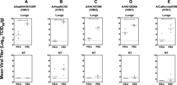
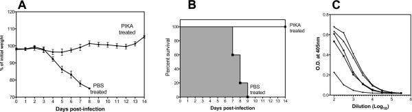
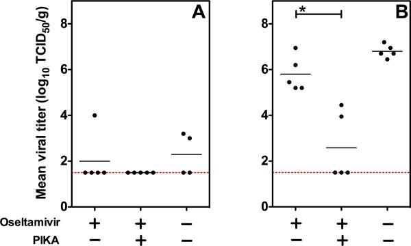
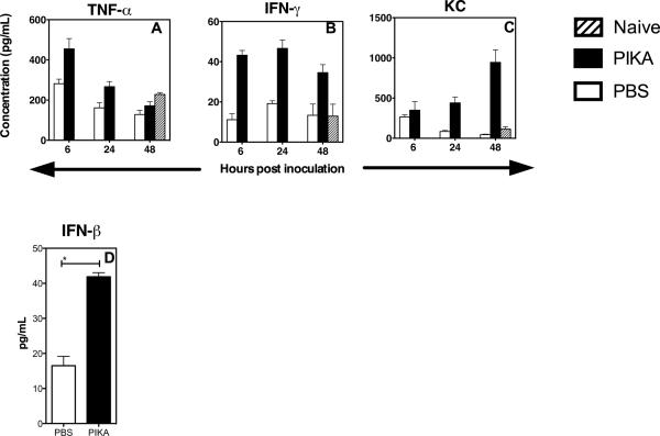
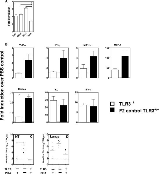
The efficacy of a stabilized chemical analog of double-stranded ribonucleic acid (RNA), PIKA, as prophylaxis against infection with 5 different influenza A virus subtypes, including the 2009 swine-origin pandemic H1N1 virus, was evaluated in mice. Intranasal treatment with PIKA resulted in a significant reduction of viral replication in the respiratory tract. The inhibitory effect was mediated by rapid infiltration of immune cells into the lungs, and production of inflammatory cytokines. While TLR3 is important for the optimal production of these inflammatory cytokines, inhibition of viral replication was still observed in TLR3(-/-) mice. In addition, a significant synergistic effect in inhibiting H5N1 virus replication was observed when PIKA was coadministered with oseltamivir. The broad-spectrum protection provided by PIKA makes it an attractive option for prophylaxis from infection with influenza A viruses.
Published by Elsevier Inc.
Figures
Similar articles
-
CLEC5A-Mediated Enhancement of the Inflammatory Response in Myeloid Cells Contributes to Influenza Virus Pathogenicity In Vivo.J Virol. 2016 Dec 16;91(1):e01813-16. doi: 10.1128/JVI.01813-16. Print 2017 Jan 1. J Virol. 2016. PMID: 27795434 Free PMC article.
-
A TLR3 ligand that exhibits potent inhibition of influenza virus replication and has strong adjuvant activity has the potential for dual applications in an influenza pandemic.Vaccine. 2009 Feb 25;27(9):1354-64. doi: 10.1016/j.vaccine.2008.12.048. Epub 2009 Jan 15. Vaccine. 2009. PMID: 19150474 Free PMC article.
-
Highly pathogenic avian influenza A H5N1 and pandemic H1N1 virus infections have different phenotypes in Toll-like receptor 3 knockout mice.J Gen Virol. 2014 Sep;95(Pt 9):1870-1879. doi: 10.1099/vir.0.066258-0. Epub 2014 May 30. J Gen Virol. 2014. PMID: 24878639 Free PMC article.
-
Modulation of Innate Immune Responses by the Influenza A NS1 and PA-X Proteins.Viruses. 2018 Dec 12;10(12):708. doi: 10.3390/v10120708. Viruses. 2018. PMID: 30545063 Free PMC article. Review.
-
Innate Immune Responses to Influenza Virus Infections in the Upper Respiratory Tract.Viruses. 2021 Oct 17;13(10):2090. doi: 10.3390/v13102090. Viruses. 2021. PMID: 34696520 Free PMC article. Review.
Cited by
-
Safety, tolerability, and immunogenicity of PIKA-adjuvanted recombinant SARS-CoV-2 spike protein subunit vaccine in healthy adults: an open-label randomized phase I clinical trial.Clin Exp Vaccine Res. 2024 Oct;13(4):315-328. doi: 10.7774/cevr.2024.13.4.315. Epub 2024 Oct 31. Clin Exp Vaccine Res. 2024. PMID: 39525677 Free PMC article.
-
TLR Agonists as Modulators of the Innate Immune Response and Their Potential as Agents Against Infectious Disease.Front Immunol. 2014 Mar 3;5:79. doi: 10.3389/fimmu.2014.00079. eCollection 2014. Front Immunol. 2014. PMID: 24624130 Free PMC article. Review.
-
Protective immunity and safety of a genetically modified influenza virus vaccine.PLoS One. 2014 Jun 13;9(6):e98685. doi: 10.1371/journal.pone.0098685. eCollection 2014. PLoS One. 2014. PMID: 24927156 Free PMC article.
-
Synergistic TLR2/6 and TLR9 activation protects mice against lethal influenza pneumonia.PLoS One. 2012;7(1):e30596. doi: 10.1371/journal.pone.0030596. Epub 2012 Jan 27. PLoS One. 2012. PMID: 22299046 Free PMC article.
-
Attenuation of Influenza A Virus Disease Severity by Viral Coinfection in a Mouse Model.J Virol. 2018 Nov 12;92(23):e00881-18. doi: 10.1128/JVI.00881-18. Print 2018 Dec 1. J Virol. 2018. PMID: 30232180 Free PMC article.
References
-
- Alexopoulou L, Holt AC, Medzhitov R, Flavell RA. Recognition of double-stranded RNA and activation of NF-kappaB by Toll-like receptor 3. Nature. 2001;413(6857):732–8. - PubMed
-
- Arimura H. Correlation between molecular size and interferon- inducing activity of poly I:C. Acta Virol. 1975;19(6):457–66. - PubMed
-
- Beigel JH, Farrar J, Han AM, Hayden FG, Hyer R, de Jong MD, Lochindarat S, Nguyen TK, Nguyen TH, Tran TH, Nicoll A, Touch S, Yuen KY. Avian influenza A (H5N1) infection in humans. N Engl J Med. 2005;353(13):1374–85. - PubMed
-
- Bernstein DI, Harrison CJ, Tomai MA, Miller RL. Daily or weekly therapy with resiquimod (R-848) reduces genital recurrences in herpes simplex virus-infected guinea pigs during and after treatment. J Infect Dis. 2001;183(6):844–9. - PubMed
Publication types
MeSH terms
Substances
Grants and funding
LinkOut - more resources
Full Text Sources
Other Literature Sources
