. 2010 Oct 10;406(1):1-11.
doi: 10.1016/j.virol.2010.07.001. Epub 2010 Jul 29.

Norwalk virus does not replicate in human macrophages or dendritic cells derived from the peripheral blood of susceptible humans

Affiliations

Norwalk virus does not replicate in human macrophages or dendritic cells derived from the peripheral blood of susceptible humans

Margarita K Lay et al. Virology. .

Abstract

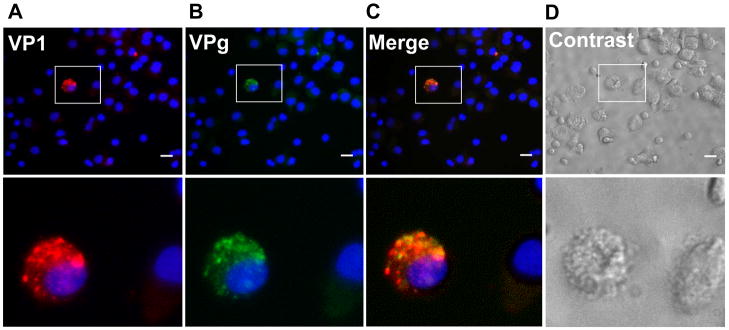
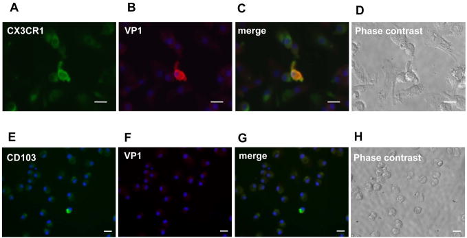
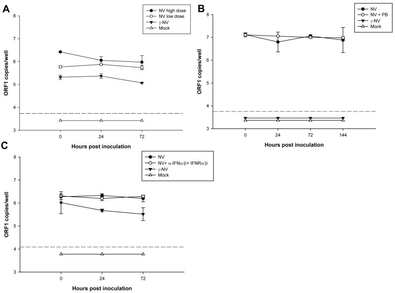
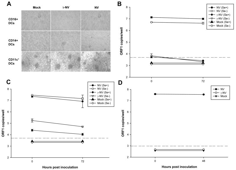
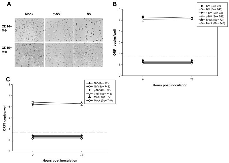
Human noroviruses are difficult to study due to the lack of an efficient in vitro cell culture system or small animal model. Murine norovirus replicates in murine macrophages (MPhi) and dendritic cells (DCs), raising the possibility that human NoVs might replicate in such human cell types. To test this hypothesis, we evaluated DCs and MPhi derived from monocyte subsets and CD11c(+) DCs isolated from peripheral blood mononuclear cells of individuals susceptible to Norwalk virus (NV) infection. These cells were exposed to NV and replication was evaluated by immunofluorescence and by quantitative RT-PCR. A few PBMC-derived DCs expressed NV proteins. However, NV RNA did not increase in any of the cells tested. These results demonstrate that NV does not replicate in human CD11c(+) DCs, monocyte-derived DCs and MPhi, but abortive infection may occur in a few DCs. These results suggest that NV tropism is distinct from that of murine noroviruses.


Figures

Figure 1
Figure 1. NV quantification in fractions from NV purified from stools by isopycnic CsCl gradient ultracentrifugation
Genomic NV RNA in each fraction of the CsCl density gradient was quantified by IMC/qRT-PCR, using primers specific for the ORF1 region of the NV genome (squares). The buoyant density of each fraction was also determined by measuring the refractive index (diamonds). Each square represents replicas of 3.
Figure 2
Figure 2. Effect of γ-irradiation on NV genomic RNA qRT-PCR detection
NV purified from stools was exposed to different γ-irradiation doses (black dots) expressed in kilo Gray units (kGy); or left unexposed at room temperature for equivalent periods of time (white dots). Quantitation of NV RNA was performed by IMC/qRT-PCR, using primers specific for the ORF1 region of the NV genome. The percentage of remaining ORF1 copy numbers is shown on a log10 scale. Lineal regression curves were calculated as r2=0.994 for γ-irradiated NV samples. Each dot represents 12 replicas.
Figure 3
Figure 3. Detection of NV proteins by immunofluorescence in a few PBMC monocyte-derived DCs inoculated with NV
NV inoculated PBMC-DCs from a type O/Se+ individual (donor 1) were dual labeled with antibodies specific for VP1 (NV capsid protein) and for VPg (NV nonstructural protein). A few cells (2–3 out of 4 x 10⁴ cells) express VP1 and VPg proteins (A, B and C). Antibodies were visualized with species-specific IgG conjugated to Alexa Fluor 594 and 488, respectively. DAPI staining specific for nucleus was also performed (A, B and C). Phase contrast image of the stained cells (D). White scale bar represents 20 μm (upper panel); an enlargement of the boxed area (lower panel).
Figure 4
Figure 4. NV protein expressing cells co-localize with some CX3CR1+ cells, but not with CD103+ cells by immunofluorescence
NV-inoculated PBMC-DCs from a type O/Se+ individual (donor 698) were stained with antibodies specific for CX3CR1 cell surface marker (A) and for capsid proteinVP1 (B). Antibodies were visualized with species-specific IgG conjugated to Alexa Fluor 488 and 594, respectively. Similar staining was performed with antibodies specific for CD103 surface marker (E) and for VP1 (F). Merge images from the respective co-staining are shown in C and G. DAPI staining specific for nucleus was also performed (A, B, C, E, F and G). Phase contrast image of the stained cells (D and H). White scale bar represents 20 μm.
Figure 5
Figure 5. Detection of genomic NV RNA in PBMC monocyte-derived DCs inoculated with NV by qRT-PCR
Total RNA combined from the cells and the supernatant of PBMC-DCs from a type O/Se+ individual (donor 1) exposed to mock (white triangles), or γ-irradiated NV (black triangles) or 275 NV genome copies/cell (white circles) or 1,800 NV genome copies/cell (black circles) of NV purified from stool, was analyzed by qRT-PCR, using primers specific to ORF1, at 0, 24 and 72 hpi (A). Total RNA from PBMC-DCs inoculated with NV compared to NV inoculated PBMC-DCs (donor 778) treated with 10 μg/ml polymixin B (B); or NV inoculated PBMC-DCs (donor 453) treated with a cocktail of antibodies against IFN-α, IFN-β and IFN-α/β receptor (C) were analyzed by qRT-PCR at different times. Total ORF1 copy numbers per well are represented on a log10 scale. Each well contained from 4–7 x10⁵ cells. The data plotted represent the means ± standard deviation of triplicate wells. Dotted lines represent the detection limit of the assay. All points below the limit of detection had the same value but were offset for visualization.
Figure 6
Figure 6. NV does not replicate in CD16+, CD14+ monocyte-derived DCs or CD11c+ blood DCs
Analysis of cell morphology of CD16+ DCs, CD14+ DCs, and CD11c+ DCs exposed to mock, γ-NV and NV after 48 hpi by phase contrast microscopy (A). The titer of NV RNA in each sample: mock, γ-NV and NV exposed CD16+ DCs (B) and CD14+ DCs (C), from type O/Se+ (donor 58) and type O/Se (donor 56) individuals; and CD11c+ DCs (D) from a type O/Se+ (donor 103) individual, was measured by qRT-PCR at 0, 48 or 72 hpi. Primers targeting the ORF1 region of the NV genomic RNA were used. Total ORF1 copy numbers per well are represented on a log10 scale. Each well contained from 2–4 x10⁵ cells. The data plotted represent the means ± standard deviation of 6 (B and C) or 4 (D) replicate wells. Dotted lines represent the detection limit of the assay. All points below the limit of detection had the same value but were offset for visualization.
Figure 7
Figure 7. NV does not replicate in CD14+ or CD16+ monocyte-derived MΦ
Analysis of cell morphology observed by phase contrast microscopy in CD14+ MΦ and CD16+ MΦ exposed to mock, γ-NV and NV after 48 hpi (A). The titer of NV RNA in each sample: mock, γ-NV and NV exposed CD14+ MΦ (B), and CD16+ MΦ (C) from type O/Se+ individuals (donors 72 and 748), was measured by qRT. PCR at 0 and 72 hpi. Primers targeting the ORF1 region of the NV genomic RNA were used. Total ORF1 copy numbers per well are represented on a log10 scale. Each well contained 2 x10⁵ cells. The data plotted represent the means ± standard deviation of 4 (B) or 3 (C) replicate wells. Dotted lines represent the detection limit of the assay. All points below the limit of detection had the same value but were offset for visualization.

Similar articles

Cited by

References

    1. Asanaka M, Atmar RL, Ruvolo V, Crawford SE, Neill FH, Estes MK. Replication and packaging of Norwalk virus RNA in cultured mammalian cells. Proc Natl Acad Sci U S A. 2005;102:10327–10332. - PMC - PubMed
    1. Atmar RL, Opekun AR, Gilger MA, Estes MK, Crawford SE, Neill FH, Graham DY. Norwalk virus shedding after experimental human infection. Emerg Infect Dis. 2008;14:1553–1557. PMC2609865. - PMC - PubMed
    1. Blutt SE, Conner ME. Rotavirus: to the gut and beyond! Curr Opin Gastroenterol. 2007;23:39–43. - PubMed
    1. Bogunovic M, Ginhoux F, Helft J, Shang L, Hashimoto D, Greter M, Liu K, Jakubzick C, Ingersoll MA, Leboeuf M, Stanley ER, Nussenzweig M, Lira SA, Randolph GJ, Merad M. Origin of the lamina propria dendritic cell network. Immunity. 2009;31:513–525. - PMC - PubMed
    1. Chang KO, George DW. Interferons and ribavirin effectively inhibit Norwalk virus replication in replicon-bearing cells. J Virol. 2007;81:12111–12118. - PMC - PubMed

Publication types

MeSH terms