. 2010 Oct 1;285(40):30567-76.
doi: 10.1074/jbc.M110.111856. Epub 2010 Jul 27.

Synergistic allostery, a sophisticated regulatory network for the control of aromatic amino acid biosynthesis in Mycobacterium tuberculosis

Affiliations

Synergistic allostery, a sophisticated regulatory network for the control of aromatic amino acid biosynthesis in Mycobacterium tuberculosis

Celia J Webby et al. J Biol Chem. .

Abstract

The shikimate pathway, responsible for aromatic amino acid biosynthesis, is required for the growth of Mycobacterium tuberculosis and is a potential drug target. The first reaction is catalyzed by 3-deoxy-d-arabino-heptulosonate 7-phosphate synthase (DAH7PS). Feedback regulation of DAH7PS activity by aromatic amino acids controls shikimate pathway flux. Whereas Mycobacterium tuberculosis DAH7PS (MtuDAH7PS) is not inhibited by the addition of Phe, Tyr, or Trp alone, combinations cause significant loss of enzyme activity. In the presence of 200 μm Phe, only 2.4 μm Trp is required to reduce enzymic activity to 50%. Reaction kinetics were analyzed in the presence of inhibitory concentrations of Trp/Phe or Trp/Tyr. In the absence of inhibitors, the enzyme follows Michaelis-Menten kinetics with respect to substrate erythrose 4-phosphate (E4P), whereas the addition of inhibitor combinations caused significant homotropic cooperativity with respect to E4P, with Hill coefficients of 3.3 (Trp/Phe) and 2.8 (Trp/Tyr). Structures of MtuDAH7PS/Trp/Phe, MtuDAH7PS/Trp, and MtuDAH7PS/Phe complexes were determined. The MtuDAH7PS/Trp/Phe homotetramer binds four Trp and six Phe molecules. Binding sites for both aromatic amino acids are formed by accessory elements to the core DAH7PS (β/α)(8) barrel that are unique to the type II DAH7PS family and contribute to the tight dimer and tetramer interfaces. A comparison of the liganded and unliganded MtuDAH7PS structures reveals changes in the interface areas associated with inhibitor binding and a small displacement of the E4P binding loop. These studies uncover a previously unrecognized mode of control for the branched pathways of aromatic amino acid biosynthesis involving synergistic inhibition by specific pairs of pathway end products.


Figures
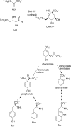
FIGURE 1.
FIGURE 1.
DAH7P synthase reaction and shikimate pathway.
FIGURE 2.
FIGURE 2.
Effect of aromatic amino acids on the reaction rate of MtuDAH7PS. A, the plot shows the effect of individual amino acids on the reaction rate of MtuDAH7PS, ranging from 0 to 200 μm. B, the plot shows the effect of a fixed amino acid concentration (200 μm) in combination with a variable amino acid concentration on MtuDAH7PS reaction rate. All data points are the means of triplicate experiments, and S.E. are shown.
FIGURE 3.
FIGURE 3.
Steady-state kinetic analysis of MtuDAH7PS. Plots showing the E4P rate dependence in the absence (black circles) and presence of a fixed concentration of aromatic amino acid (A), and combinations of aromatic amino acids (B). The data were fitted to the Michaelis-Menten equation and the Hill equation for cooperativity that described the sigmoidal behavior seen in the presence of Trp in the combinations plot.
FIGURE 4.
FIGURE 4.
a, the homotetramer of MtuDAH7PS in complex with Phe and Trp is shown. Ligand molecules are displayed in green. b, the monomer structure of MtuDAH7PS is shown. In both panels, the N-terminal extra-barrel elements (the N termini and α0a-α0c helices) are shown in red, and the additional α2a, α2b helices are shown in yellow. Active site metal ions are shown as purple spheres, and the ligands are in sphere form. The Phe binding in secondary site is shown with orange carbon atoms, and the Phe in primary site and the Trp ligand are shown with green carbons.
FIGURE 5.
FIGURE 5.
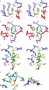
Ligand binding sites and active site. a, shown are the primary Phe binding sites in the tight dimer interface. Residues 3–10 are excluded for clarity. b, shown is the secondary Phe binding site in subunit B. c, shown are the Trp binding sites. In all three figures residues on the basic barrel structure are shown with blue carbons, residues that are on the N-terminal extra-barrel addition are shown in red, and the residues on the α2a-α2b helices are shown with yellow carbons. The bound Trp and Phe molecules are shown with green carbons. Water molecules are displayed as red spheres. Hydrogen bonds are displayed as red dashed lines. d, superposition of the active site of the Trp+Phe structure and the unliganded Thesit-free structure of MtuDAH7PS. Residues from the Thesit-free unliganded structure are shown with cyan carbons, and residues from the Trp+Phe structure are shown with green carbons. The PEP bound in the active site of molecule A in the Thesit-free structure is shown in yellow for comparison with the position of the phosphate ion from the Trp+Phe structure (shown in red). The side chains of residues 134–139 are omitted for clarity. The alternative backbone position of residues 135–139 is shown with purple carbons.
FIGURE 6.
FIGURE 6.
The individual and combined effects of added amino acids on the reaction rate of MtuDAH7PS and amino acid binding site mutants. The specific activities for each protein are shown in the absence of amino acids and in the presence of 200 μm Trp, 200 μm Phe, and 200 μm Trp and Phe in combination. All data points are the means of triplicate experiments, and S.E. are shown. The R171A and L194A mutants and R256A mutants target the primary Phe, the Trp, and secondary Phe binding sites, respectively.

References

    1. Bentley R. (1990) Crit. Rev. Biochem. Mol. Biol. 25, 307–384 - PubMed
    1. Walsh C. T., Liu J., Rusnak F., Sakaitani M. (1990) Chem. Rev. 90, 1105–1129
    1. Roberts F., Roberts C. W., Johnson J. J., Kyle D. E., Krell T., Coggins J. R., Coombs G. H., Milhous W. K., Tzipori S., Ferguson D. J., Chakrabarti D., McLeod R. (1998) Nature 393, 801–805 - PubMed
    1. Campbell S. A., Richards T. A., Mui E. J., Samuel B. U., Coggins J. R., McLeod R., Roberts C. W. (2004) Int. J. Parasitol. 34, 5–13 - PubMed
    1. Parish T., Stoker N. G. (2002) Microbiology 148, 3069–3077 - PubMed

Publication types

MeSH terms

LinkOut - more resources