Membrane uncoating of intact enveloped viruses
- PMID: 20668081
- PMCID: PMC2953184
- DOI: 10.1128/JVI.00229-10
Membrane uncoating of intact enveloped viruses
Abstract
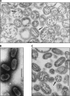
Experiments in the 1960s showed that Sendai virus, a paramyxovirus, fused its membrane with the host plasma membrane. After membrane fusion, the virus spontaneously "uncoated" with diffusion of the viral membrane proteins into the host plasma membrane and a merging of the host and viral membranes. This led to deposit of the viral ribonucleoprotein (RNP) and interior proteins in the cell cytoplasm. Later work showed that the common procedure then used to grow Sendai virus produced damaged, pleomorphic virions. Virions, which were grown under conditions that were not damaging, made a connecting structure between virus and cell at the region where the fusion occurred. The virus did not release its membrane proteins into the host membrane. The viral RNP was seen in the connecting structure in some cases. Uncoating of intact Sendai virus proceeds differently from uncoating described by the current standard model developed long ago with damaged virus. A model of intact paramyxovirus uncoating is presented and compared to what is known about the uncoating of other viruses.
Figures


Similar articles
-
The Viral Polymerase Complex Mediates the Interaction of Viral Ribonucleoprotein Complexes with Recycling Endosomes during Sendai Virus Assembly.mBio. 2020 Aug 25;11(4):e02028-20. doi: 10.1128/mBio.02028-20. mBio. 2020. PMID: 32843550 Free PMC article.
-
The Art of Viral Membrane Fusion and Penetration.Subcell Biochem. 2023;106:113-152. doi: 10.1007/978-3-031-40086-5_4. Subcell Biochem. 2023. PMID: 38159225
-
A spatio-temporal analysis of matrix protein and nucleocapsid trafficking during vesicular stomatitis virus uncoating.PLoS Pathog. 2010 Jul 15;6(7):e1000994. doi: 10.1371/journal.ppat.1000994. PLoS Pathog. 2010. PMID: 20657818 Free PMC article.
-
Membrane fusion of enveloped viruses: especially a matter of proteins.J Bioenerg Biomembr. 1990 Apr;22(2):121-55. doi: 10.1007/BF00762943. J Bioenerg Biomembr. 1990. PMID: 2109749 Review.
-
[Envelope virus assembly and budding].Uirusu. 2010 Jun;60(1):105-13. doi: 10.2222/jsv.60.105. Uirusu. 2010. PMID: 20848870 Review. Japanese.
Cited by
-
Structural analysis of respiratory syncytial virus reveals the position of M2-1 between the matrix protein and the ribonucleoprotein complex.J Virol. 2014 Jul;88(13):7602-17. doi: 10.1128/JVI.00256-14. Epub 2014 Apr 23. J Virol. 2014. PMID: 24760890 Free PMC article.
-
Human hepatoma HepG2 cells are susceptible to infection by domestic cat hepadnavirus.J Vet Diagn Invest. 2025 Jul;37(4):609-619. doi: 10.1177/10406387251339765. Epub 2025 May 12. J Vet Diagn Invest. 2025. PMID: 40353562 Free PMC article.
-
The Dynamic Landscape of Capsid Proteins and Viral RNA Interactions in Flavivirus Genome Packaging and Virus Assembly.Pathogens. 2024 Jan 28;13(2):120. doi: 10.3390/pathogens13020120. Pathogens. 2024. PMID: 38392858 Free PMC article. Review.
-
Alphavirus genome delivery occurs directly at the plasma membrane in a time- and temperature-dependent process.J Virol. 2013 Apr;87(8):4352-9. doi: 10.1128/JVI.03412-12. Epub 2013 Feb 6. J Virol. 2013. PMID: 23388718 Free PMC article.
-
Pseudotyped Viruses for Retroviruses.Adv Exp Med Biol. 2023;1407:61-84. doi: 10.1007/978-981-99-0113-5_4. Adv Exp Med Biol. 2023. PMID: 36920692
References
-
- Bashford, C. L., G. M. Alder, G. Menestrina, K. J. Micklem, J. J. Murphy, and C. A. Pasternak. 1986. Membrane damage by hemolytic viruses, toxins, complement, and other cytotoxic agents: a common mechanism blocked by divalent cations. J. Biol. Chem. 261:9300-9308. - PubMed
-
- Bittman, R., Z. Majuk, D. S. Honig, R. W. Compans, and J. Lenard. 1976. Permeability properties of the membrane of vesicular stomatitis virions. Biochim. Biophys. Acta 433:63-74. - PubMed
-
- Branton, D., S. Bullivant, N. B. Gilula, M. J. Karnovsky, H. Moor, K. Mühlethaler, D. H. Northcote, L. Packer, B. Satir, P. Satir, V. Speth, L. A. Staehlin, R. L. Steere, and R. S. Weinstein. 1975. Freeze-etching nomenclature. Science 190:54-56. - PubMed
Publication types
MeSH terms
Substances
LinkOut - more resources
Full Text Sources

