. 2010 Oct;84(19):10102-12.
doi: 10.1128/JVI.01208-10. Epub 2010 Jul 28.

Proteolytic products of the porcine reproductive and respiratory syndrome virus nsp2 replicase protein

Affiliations

Proteolytic products of the porcine reproductive and respiratory syndrome virus nsp2 replicase protein

Jun Han et al. J Virol. 2010 Oct.

Abstract

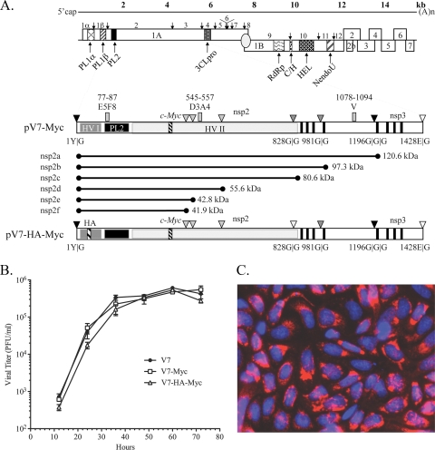
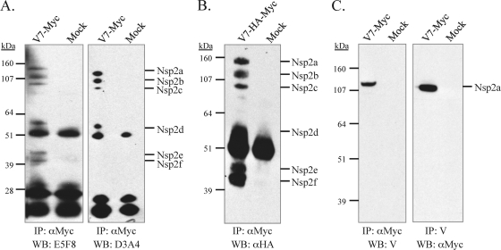
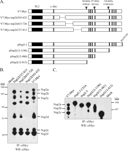
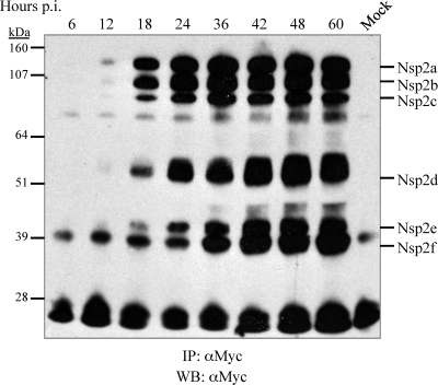
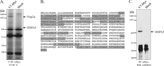
The nsp2 replicase protein of porcine reproductive and respiratory syndrome virus (PRRSV) was recently demonstrated to be processed from its precursor by the PL2 protease at or near the G(1196)|G(1197) dipeptide in transfected CHO cells. Here the proteolytic cleavage of PRRSV nsp2 was further investigated in virally infected MARC-145 cells by using two recombinant PRRSVs expressing epitope-tagged nsp2. The data revealed that PRRSV nsp2 exists as different isoforms, termed nsp2a, nsp2b, nsp2c, nsp2d, nsp2e, and nsp2f, during PRRSV infection. Moreover, on the basis of deletion mutagenesis and antibody probing, these nsp2 species appeared to share the same N terminus but to differ in their C termini. The largest protein, nsp2a, corresponded to the nsp2 product identified in transfected CHO cells. nsp2b and nsp2c were processed within or near the transmembrane (TM) region, presumably at or near the conserved sites G(981)|G(982) and G(828)|G(829)|G(830), respectively. The C termini for nsp2d, -e, and -f were mapped within the nsp2 middle hypervariable region, but no conserved cleavage sites could be definitively predicted. The larger nsp2 species emerged almost simultaneously in the early stage of PRRSV infection. Pulse-chase analysis revealed that all six nsp2 species were relatively stable and had low turnover rates. Deletion mutagenesis revealed that the smaller nsp2 species (e.g., nsp2d, nsp2e, and nsp2f) were not essential for viral replication in cell culture. Lastly, we identified a cellular chaperone, named heat shock 70-kDa protein 5 (HSPA5), that was strongly associated with nsp2, which may have important implications for PRRSV replication. Overall, these findings indicate that PRRSV nsp2 is increasingly emerging as a multifunctional protein and may have a profound impact on PRRSV replication and viral pathogenesis.


Figures

FIG. 1.
FIG. 1.
Characterization of foreign-epitope-tagged PRRSV. (A) PRRSV genome annotation. The genome of PRRSV is shown with all identified open reading frames. ORF1A and ORF1B are posttranslationally cleaved by virally encoded papain-like proteases (PCP1α and PCP1β), a cysteine protease (PL2), and a poliovirus 3C-like serine protease (3CL). The following polymerase signature regions are indicated: RNA-dependent RNA polymerase (RdRp), cysteine and histidine rich (C/H), helicase (HEL), and nidovirus uridylate-specific endoribonuclease (NendoU). Below the genome schematic are expanded diagrams of nsp2-3 and the construction of recombinant PRRSVs expressing c-myc- or c-myc- and HA-tagged nsp2. nsp2 contains an N-terminal small hypervariable region (HV-I), a PL2 protease domain, a middle hypervariable region (HV-II), a putative transmembrane domain (filled vertical bars), and a C-terminal domain. The nsp2 PL2 protease is predicted to cleave at G|G dipeptides; there are 10 such dipeptides in type 2 strain VR-2332, some of which are not conserved in other strains. The major conserved predicted cleavage sites discussed in this report are represented by dark shaded triangles; potential cleavage sites that are not conserved are shown as light shaded triangles; and the cleavage sites of nsp1|nsp2 and nsp3|nsp4 are shown as filled and open triangles, respectively. Three consecutive c-myc epitopes were inserted in place of nsp2 aa 324 to 434, based on strain VR-2332 full-length cDNA clone pVR-V7, to generate mutant pV7-myc. In the bottom construct, an HA epitope replaced nsp2 aa 12 to 24 of pV7-myc to generate the double-tagged mutant pV7-HA-myc. (B) Growth kinetics of mutants V7-myc and V7-HA-myc and of parental virus VR-V7 at passage 3. The viruses were used to infect MARC-145 cells in T25 flasks at an MOI of 0.1. The virus-infected cell supernatants were collected every 12 h and were titrated by a viral plaque assay. The mean results were plotted; error bars indicate standard deviations. (C) Immunostaining of nsp2 protein. At 20 h postinfection, the nsp2 protein in V7-myc-infected MARC-145 cells was labeled using monoclonal antibody 9E10 against a c-myc epitope and an Alexa 568-conjugated secondary antibody (red) (Molecular Probes). Nuclei were stained with DAPI (blue). The fields were merged using Photoshop, version 8.
FIG. 2.
FIG. 2.
Identification of nsp2 products in PRRSV-infected MARC-145 cells. (A) MARC-145 cells either were mock infected or were infected with V7-myc (MOI, 0.1) at passage 3. At 24 to 36 h postinfection, the cells were lysed, and nsp2 proteins were immunoprecipitated (IP) by mouse monoclonal antibody 9E10 recognizing the c-myc epitope. The samples were analyzed by reducing SDS-PAGE on a 4 to 12% polyacrylamide gel followed by Western blotting (WB) with the nsp2-specific antibody E5F8 or D3A4. (B) Analysis of nsp2-associated products in V7-HA-myc-infected MARC-145 cells. The nsp2 products were first immunoprecipitated by anti-c-myc antibody 9E10, then separated by SDS-PAGE, and finally analyzed by WB with a mouse monoclonal antibody to the HA epitope. (C) V7-myc-infected cell lysates were immunoprecipitated with anti-c-myc antibody 9E10 or rabbit antibody V, recognizing a peptide near the C terminus of nsp2; then they were separated by SDS-PAGE and analyzed by immunoblotting with antibody V or rabbit anti-c-myc polyclonal antibodies. The use of different antibody combinations for IP and WB frequently detected nonspecific proteins (25 kDa and 50 kDa, respectively), which are likely immunoglobulin light and heavy chains, respectively, that reacted with the secondary antibody.
FIG. 3.
FIG. 3.
Mapping of the relative positions of the nsp2 isoforms. (A) V7-myc is shown in schematic form with PL2 cleavage sites and the calculated molecular sizes of predicted proteins that included the nsp2 N terminus. V7-myc nsp2 regions comprising aa 543 to 632, aa 633 to 726, or aa 727 to 813 were deleted to generate new full-length infectious cDNA clone mutants pV7-myc-nsp2Δ543-632, pV7-myc-nsp2Δ633-726, and pV7-myc-nsp2Δ727-813, respectively. Polypeptides corresponding to nsp2 aa 12 to 813, aa 12 to 981, and aa 12 to 1196 were cloned into pcDNA3 to generate plasmid constructs pNsp2(12-813), pNsp2(12-981), and pNsp2(12-1196). The HA-FLAG epitope was attached to the C terminus of each polypeptide. The predicted molecular sizes for the corresponding shortened polypeptides were calculated and are given in the text. (B) MARC-145 cells were infected with nsp2 deletion mutant V7-myc-nsp2Δ543-632, V7-myc-nsp2Δ633-726, or V7-myc-nsp2Δ727-813. At 24 to 36 h postinfection, the cells were lysed and immunoprecipitated (IP) with the anti-c-myc monoclonal antibody 9E10, separated by SDS-PAGE on a 4 to 12% NuPage gel, and subjected to Western blotting (WB) with anti-c-myc rabbit polyclonal antibodies. (C) CHO cells were transfected with plasmids expressing either nsp2-3, nsp2-3(C55A), which does not undergo PL2 proteolysis (14), or one of the nsp2 truncation mutants. The cells were lysed after 48 h of transfection; then they were immunoprecipitated with anti-c-myc monoclonal antibody 9E10 and analyzed by Western blotting with an anti-c-myc rabbit polyclonal antibody. The nsp2 proteins immunoprecipitated from V7-myc-infected MARC-145 cells served as a control. The extra bands in the lanes for truncation mutants Nsp2(12-1196) and Nsp2(12-981) may represent degraded products, possibly due to cellular proteases.
FIG. 4.
FIG. 4.
Accumulation of the nsp2 isoforms during virus infection. MARC-145 cells in 60-mm-diameter petri dishes were infected with V7-myc at an MOI of 0.1. At different time points after infection, as indicated, the cells were lysed, immunoprecipitated (IP) with monoclonal antibody 9E10 against the c-myc epitope, separated by SDS-PAGE on a 4 to 12% NuPage gel with 5% β-mercaptoethanol, and subjected to Western blotting (WB) with rabbit polyclonal antibodies to c-myc.
FIG. 5.
FIG. 5.
Pulse-chase analysis of the nsp2 isoforms. (A to D) MARC-145 cells in 60-mm-diameter petri dishes were infected with V7-myc at an MOI of 0.1. At 20 h postinfection, the cells were labeled with [35S]methionine-cysteine. Cells were lysed and immunoprecipitated (IP) with either anti-c-myc antibody 9E10 (A) or rabbit antipeptide antibody V (C). For pulse-chase analysis, the cells were pulsed for 5 h and then chased for as long as 4 h. nsp2 proteins were immunoprecipitated with anti-c-myc monoclonal antibody 9E10 (B) or rabbit antipeptide antibody V (D). (E and F) In an effort to elucidate the precursor-product relationship, cells were pulsed for 20 min and chased for as long as 2 h. nsp2 proteins were then immunoprecipitated with anti-c-myc monoclonal antibody 9E10 (E) or rabbit antipeptide antibody V (F). The proteins were separated by 4 to 12% SDS-PAGE with 10% dithiothreitol. The gel was dried, and radiolabeled nsp2 proteins were detected by autoradiography.
FIG. 6.
FIG. 6.
Coimmunoprecipitation of HSPA5 with nsp2. (A) MARC-145 cells were infected with V7-myc at an MOI of 0.1. At 24 to 36 h postinfection, the cells were lysed and subjected to two rounds of immunoprecipitation (IP), with anti-c-myc monoclonal antibody 9E10 and rabbit antipeptide antibody V, respectively. The proteins were resolved on a 4 to 12% NuPage reducing gel and were stained with Coomassie blue. (B) The separated proteins were then stained with Sypro Ruby, excised, digested with trypsin, extracted, and subjected to LC-MS-MS analysis. The HSPA5 (GRP78) amino acid sequence of Macaca mulatta (rhesus monkey; GenBank ID 109112231) is shown, with the recovered sequences, sometimes overlapping each other, highlighted in gray. (C) Immunoblot analysis of the immunoprecipitated proteins with a rabbit polyclonal antibody to HSPA5.

Similar articles

Cited by

References

    1. Carter, K. L., and B. Roizman. 1996. The promoter and transcriptional unit of a novel herpes simplex virus 1 alpha gene are contained in, and encode a protein in frame with, the open reading frame of the alpha 22 gene. J. Virol. 70:172-178. - PMC - PubMed
    1. Castón, J. R., J. L. Martinez-Torrecuadrada, A. Maraver, E. Lombardo, J. F. Rodriguez, J. I. Casal, and J. L. Carrascosa. 2001. C terminus of infectious bursal disease virus major capsid protein VP2 is involved in definition of the T number for capsid assembly. J. Virol. 75:10815-10828. - PMC - PubMed
    1. Cavanagh, D. 1997. Nidovirales: a new order comprising Coronaviridae and Arteriviridae. Arch. Virol. 142:629-633. - PubMed
    1. Cho, D. Y., G. H. Yang, C. J. Ryu, and H. J. Hong. 2003. Molecular chaperone GRP78/BiP interacts with the large surface protein of hepatitis B virus in vitro and in vivo. J. Virol. 77:2784-2788. - PMC - PubMed
    1. Choukhi, A., S. Ung, C. Wychowski, and J. Dubuisson. 1998. Involvement of endoplasmic reticulum chaperones in the folding of hepatitis C virus glycoproteins. J. Virol. 72:3851-3858. - PMC - PubMed

Publication types

MeSH terms

LinkOut - more resources