. 2010 Oct;84(19):10004-15.
doi: 10.1128/JVI.01151-10. Epub 2010 Jul 28.

Interplay of acute and persistent infections caused by Venezuelan equine encephalitis virus encoding mutated capsid protein

Affiliations

Interplay of acute and persistent infections caused by Venezuelan equine encephalitis virus encoding mutated capsid protein

Svetlana Atasheva et al. J Virol. 2010 Oct.

Abstract

Venezuelan equine encephalitis virus (VEEV) is a significant human and animal pathogen. The highlight of VEEV replication in vitro, in cells of vertebrate origin, is the rapid development of cytopathic effect (CPE), which is strongly dependent upon the expression of viral capsid protein. Besides being an integral part of virions, the latter protein is capable of (i) binding both the nuclear import and nuclear export receptors, (ii) accumulating in the nuclear pore complexes, (iii) inhibiting nucleocytoplasmic trafficking, and (iv) inhibiting transcription of cellular ribosomal and messenger RNAs. Using our knowledge of the mechanism of VEEV capsid protein function in these processes, we designed VEEV variants containing combinations of mutations in the capsid-coding sequences. These mutations made VEEV dramatically less cytopathic but had no effect on infectious virus production. In cell lines that have defects in type I interferon (IFN) signaling, the capsid mutants demonstrated very efficient persistent replication. In other cells, which have no defects in IFN production or signaling, the same mutants were capable of inducing a long-term antiviral state, downregulating virus replication to an almost undetectable level. However, ultimately, these cells also developed a persistent infection, characterized by continuous virus replication and beta IFN (IFN-beta) release. The results of this study demonstrate that the long-term cellular antiviral state is determined by the synergistic effects of type I IFN signaling and the antiviral reaction induced by replicating viral RNA and/or the expression of VEEV-specific proteins. The designed mutants represent an important model for studying the mechanisms of cell interference with VEEV replication and development of persistent infection.


Figures

FIG. 1.
FIG. 1.
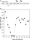
Schematic representation of recombinant VEEV TC-83 genomes having mutations in the H68 peptide of capsid protein, the infectivity of in vitro-synthesized viral RNAs, and virus titers at 24 after transfection of BHK-21 cells. SG indicates positions of the subgenomic promoters. Point mutations introduced into capsid protein are indicated by lowercase red letters. Boxes indicate nuclear export (NES) and nuclear import (NLS) signals. The connective peptide sequence is underlined.
FIG. 2.
FIG. 2.
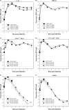
Replication of capsid mutant in the indicated cell lines. Cells were infected at an MOI of 20 PFU/cell, media were replaced at the indicated times, and virus titers were determined by a plaque assay using BHK-21 cells. Dashed lines indicate the limits of detection.
FIG. 3.
FIG. 3.
VEEV/C1/GFP develops persistent replication in STAT1−/− and IFN-α/βR−/− MEFs, but its replication is strongly inhibited in wt MEFs and NIH 3T3 cells. Cells (5 × 10⁵) were infected with the VEEV/C1/GFP variant at an MOI of 20 PFU/cell. Images of the cells were acquired at day 6 postinfection with an inverted Nikon fluorescence microscope. The same exposure times were used for detection of GFP-positive cells.
FIG. 4.
FIG. 4.
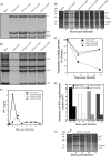
Analysis of VEEV capsid mutants replication in the NIH 3T3 cells. (A) NIH 3T3 cells were infected with the indicated viruses at an MOI of 20 PFU/cell. Between 3 and 8 h postinfection, virus-specific RNAs were metabolically labeled with [3H]uridine in the presence of ActD as described in Materials and Methods and analyzed by agarose gel electrophoresis followed by autoradiography. (B) NIH 3T3 cells were infected as described above, and between 21 and 24 h postinfection, RNAs were metabolically labeled with [3H]uridine in the absence of ActD. Isolated RNAs were analyzed by gel electrophoresis followed by autoradiography. (C) NIH 3T3 cells were infected as described above. At the indicated time points, media were replaced and concentrations of released IFN-β were determined by ELISA. (D) NIH 3T3 cells were infected at an MOI of 20 PFU/cell. At the indicated time points, they were metabolically labeled with [35S]methionine for 30 min at 37°C, and equal amounts of cell lysates were analyzed by SDS-PAGE followed by autoradiography. The same gel was also analyzed with a phosphorimager to assess synthesis of cellular (E) and viral structural (F) proteins. (G) NIH 3T3 cells were infected with VEEV/C1/GFP at an MOI of 20 PFU/cell. At the indicated time points, they were metabolically labeled with [35S]methionine for 30 min at 37°C, and equal amounts of cell lysates were analyzed by SDS-PAGE followed by autoradiography. All of the presented experiments have been repeated at least twice, and one of the highly reproducible results is presented. Mock, mock-infected cells.
FIG. 5.
FIG. 5.
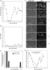
Analysis of VEEV/C1/GFP replication and IFN-β induction in NIH 3T3 cells. NIH 3T3 cells (2.5 × 10⁶) were infected with the VEEV/C1/GFP variant at an MOI of 20 PFU/cell. At the indicated time points, media were harvested, and virus titers (A) and the concentrations of IFN-β (B) were determined by plaque assay and ELISA, respectively (see Materials and Methods for details). At the same time points, cells were split at a 1:2 ratio. (C) Images of the cells were acquired before cell splitting at different times postinfection (PI) with an inverted Nikon fluorescence microscope. (D) During splitting, one-tenth of cells was harvested, and cell lysates were analyzed by Western blotting using VEEV nsP2 and VEEV capsid-specific antibodies, followed by quantitative analysis using a LICOR imager. (E) At the indicated time points, concentrations of viral genomic and subgenomic RNAs were measured by RT-qPCR as described in Materials and Methods.
FIG. 6.
FIG. 6.
Analysis of replication of superinfecting viruses VEEV TC-83, SINV Toto1101, and VSV in NIH 3T3 cells persistently infected with VEEV/C1/GFP. (A) Titers of the indicated viruses at 48 h postinfection of the naïve NIH 3T3 cells at an MOI of 20 PFU/cell. (B) NIH 3T3 cells were infected with VEEV/C1/GFP in the experiment presented in Fig. 4 and superinfected with the indicated viruses at an MOI of 20 PFU/cell. Medium was added to the cells without removing the inoculum. Titers were measured following a 48-h incubation at 37°C. (C) NIH 3T3 cells were treated with IFN-β at a concentration of 1,000 IU/ml, corresponding to IFN-β concentrations detected in the medium of the NIH 3T3 cells at 24 h postinfection with VEEV/C1/GFP (see Fig. 2 and 4 for details). After 24 h of treatment, media were replaced by those containing no IFN, and at the indicated time points, cells were infected with the viruses at an MOI of 20 PFU/ml. Titers of the released viruses were determined at 24 h postinfection by using a plaque assay on BHK-21 cells.
FIG. 7.
FIG. 7.
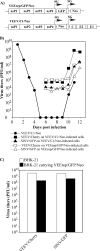
Analysis of VEEV, SINV, and VSV replication in the cells carrying VEEV replicons or infected with VEEV capsid mutant. (A) The schematic representation of VEErep/GFP/Neo replicon and VEEV/C1/Neo viral genomes. (B) NIH 3T3 cells were infected with packaged replicons and virus at MOIs of 20 inf.u/cell and 20 PFU/cell, respectively. G418 selection was started at 6 h postinfection, and cells were superinfected with VEEV/Cherry or SINV/GFP at an MOI of 20 PFU/cell. Viruses were harvested after a 48-h incubation at 37°C. Titers were determined by a plaque assay on BHK-21 cells. VEEV/Cherry and SINV/GFP titers corresponding to 105 and 104 PFU/ml, respectively, represent the residual viruses used for infection, as tested in additional experiments (data not shown). (C) BHK-21 cells were infected with the packaged VEErep/Neo/GFP replicon at an MOI of 20 inf.u/cell and, at 48 h after primary infection, superinfected with VEEV/Cherry or SINV/GFP at an MOI of 20 PFU/cell. The same numbers of naïve BHK-21 cells were infected as controls. Viruses were harvested at 48 h postinfection, and titers were determined by plaque assay on BHK-21 cells. The experiments were repeated with the indicated constructs and other viruses four times and produced very similar, reproducible results.
FIG. 8.
FIG. 8.
Analysis of SINV/G/GFP replication in the NIH 3T3 cells. (A) The schematic representation of SINV/G/GFP genome having the previously described (11) P726→G mutation in nsP2. (B) NIH 3T3 cells (2.5 × 10⁶) were infected with the SINV/G/GFP variant at an MOI of 20 PFU/cell. At the indicated time points, media were harvested, and virus titers were determined by plaque assay on BHK-21 cells. At the same time points, cells were trypsinized and split at a 1:2 ratio.

Similar articles

Cited by

References

    1. Aguilar, P. V., S. C. Weaver, and C. F. Basler. 2007. Capsid protein of eastern equine encephalitis virus inhibits host cell gene expression. J. Virol. 81:3866-3876. - PMC - PubMed
    1. Amor, S., M. F. Scallan, M. M. Morris, H. Dyson, and J. K. Fazakerley. 1996. Role of immune responses in protection and pathogenesis during Semliki Forest virus encephalitis. J. Gen. Virol. 77:281-291. - PubMed
    1. Atasheva, S., A. Fish, M. Fornerod, and E. I. Frolova. 2010. Venezuelan equine encephalitis virus capsid protein forms a tetrameric complex with CRM1 and importin α/β that obstructs nuclear pore complex function. J. Virol. 84:4158-4171. - PMC - PubMed
    1. Atasheva, S., N. Garmashova, I. Frolov, and E. Frolova. 2008. Venezuelan equine encephalitis virus capsid protein inhibits nuclear import in mammalian but not in mosquito cells. J. Virol. 82:4028-4041. - PMC - PubMed
    1. Borgherini, G., P. Poubeau, A. Jossaume, A. Gouix, L. Cotte, A. Michault, C. Arvin-Berod, and F. Paganin. 2008. Persistent arthralgia associated with chikungunya virus: a study of 88 adult patients on Reunion Island. Clin. Infect. Dis. 47:469-475. - PubMed

Publication types

MeSH terms