. 2010 Jul 28;30(30):9984-9.
doi: 10.1523/JNEUROSCI.1223-10.2010.

Persistence of coordinated long-term potentiation and dendritic spine enlargement at mature hippocampal CA1 synapses requires N-cadherin

Affiliations

Persistence of coordinated long-term potentiation and dendritic spine enlargement at mature hippocampal CA1 synapses requires N-cadherin

Ozlem Bozdagi et al. J Neurosci. .

Abstract

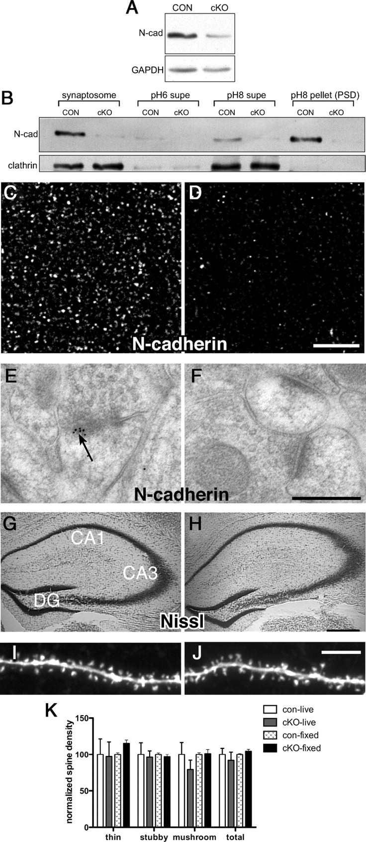
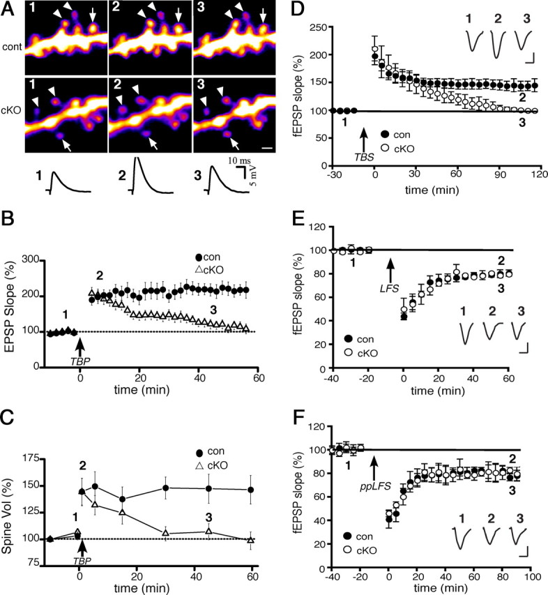
Persistent changes in spine shape are coupled to long-lasting synaptic plasticity in hippocampus. The molecules that coordinate such persistent structural and functional plasticity are unknown. Here, we generated mice in which the cell adhesion molecule N-cadherin was conditionally ablated from postnatal, excitatory synapses in hippocampus. We applied to adult mice of either sex a combination of whole-cell recording, two-photon microscopy, and spine morphometric analysis to show that postnatal ablation of N-cadherin has profound effects on the stability of coordinated spine enlargement and long-term potentiation (LTP) at mature CA1 synapses, with no effects on baseline spine density or morphology, baseline properties of synaptic neurotransmission, or long-term depression. Thus, N-cadherin couples persistent spine structural modifications with long-lasting synaptic functional modifications associated selectively with LTP, revealing unexpectedly distinct roles at mature synapses in comparison with earlier, broader functions in synapse and spine development.


Figures

Figure 1.
Figure 1.
N-cadherin is conditionally deleted from adult CA1 synapses without affecting density or morphology of dendritic spines. A, Representative immunoblot of CA1 lysates from cKO and control mice. B, Synaptosome and Triton-soluble [pH 6 supernatant (supe), containing vesicles and nonsynaptic membrane, and pH 8 supe, enriched with presynaptic matrix and junctional proteins] and -insoluble (pH 8; PSD) fractions. Equal amounts of protein were loaded in each lane and immunolabeled for N-cadherin. C, D, Immunofluorescent localization of N-cadherin in control (C) or cKO (D) CA1 stratum radiatum. Representative images were acquired under identical acquisition parameters. Scale bar, 5 μm. E, F, Postembedding immunogold labeling in CA1 shows localization of N-cadherin at control synapses (arrow, E), but an absence of gold labeling at cKO synapses (F). Scale bar, 300 nm. G, H, Nissl-staining shows no differences in hippocampal cytoarchitecture between controls (G) and cKOs (H) as expected. Scale bar, 200 μm. I, J, Images of spine-bearing apical dendrites of control (I) or cKO (J) CA1 pyramidal neurons filled live in acutely prepared hippocampal slices. Scale bar, 5 μm. K, Quantitative analysis shows no differences between genotypes in overall spine density, nor in density of morphological subtypes (thin, stubby, or mushroom spines). Spine analysis was conducted on cells filled in living slices (live) or in fixed slices (fixed). For each condition, data are normalized to control values (for further analysis, see supplemental Fig. 3, available at www.jneurosci.org).
Figure 2.
Figure 2.
LTP and spine expansion are transient, but LTD is normal, in adult cKO neurons. A, Representative images and EPSP traces from single CA1 neurons showing persistent spine expansion with the induction of TBP-LTP in control neurons (top row) but transient expansion (bottom row) and potentiation (traces) in cKO neurons. Expanded and stable spines are marked by arrowheads and arrows, respectively. The spines and EPSP traces are shown before (1), 5 min after (2), and 45 min after (3) TBP, and correspond to times shown in the population data (B, C). Scale bar, 1 μm. B, Population data showing EPSPs recorded from the same neurons as in C. TBP (arrow) induces only a transient potentiation in cKO neurons (open) but long-lasting LTP in control neurons. C, Population data showing spine enlargement following TBP (arrow) in cKO or control neurons. Initial spine expansion in the cKO neurons is identical to controls, but spine expansion becomes unstable without N-cadherin. Data shown in B and C were collected from a total of 70 spines from 12 cells from 12 slices (cKOs), and a total of 64 spines from 11 cells from 11 slices (controls). D, Field recordings of TBS-induced LTP (arrow) in CA1 in acute slices (n = 12 cKO slices; 11 control slices) taken from the same cKO and control mice shown in A–C. Inset, In this and subsequent panels, representative EPSP traces recorded at times indicated. Calibration: 10 ms, 0.5 mV. E, F, Field recordings in CA1 in acute slices from adult cKO or control mice show no differences between genotypes in two kinds of LTD [LFS, an NMDA receptor-dependent form (E), or ppLFS, a metabotropic receptor, protein synthesis-dependent form (F)].

References

    1. Bamji SX, Shimazu K, Kimes N, Huelsken J, Birchmeier W, Lu B, Reichardt LF. Role of beta-catenin in synaptic vesicle localization and presynaptic assembly. Neuron. 2003;40:719–731. - PMC - PubMed
    1. Bozdagi O, Shan W, Tanaka H, Benson DL, Huntley GW. Increasing numbers of synaptic puncta during late-phase LTP: N-cadherin is synthesized, recruited to synaptic sites, and required for potentiation. Neuron. 2000;28:245–259. - PubMed
    1. Bozdagi O, Valcin M, Poskanzer K, Tanaka H, Benson DL. Temporally distinct demands for classic cadherins in synapse formation and maturation. Mol Cell Neurosci. 2004;27:509–521. - PMC - PubMed
    1. Bozdagi O, Rich E, Tronel S, Sadahiro M, Patterson K, Shapiro ML, Alberini CM, Huntley GW, Salton SR. The neurotrophin-inducible gene Vgf regulates hippocampal function and behavior through a brain-derived neurotrophic factor-dependent mechanism. J Neurosci. 2008;28:9857–9869. - PMC - PubMed
    1. Brock JH, Elste A, Huntley GW. Distribution and injury-induced plasticity of cadherins in relationship to identified synaptic circuitry in adult rat spinal cord. J Neurosci. 2004;24:8806–8817. - PMC - PubMed

Publication types

MeSH terms