

Multiple mechanisms of consciousness: the neural correlates of emotional awareness
- PMID: 20668188
- PMCID: PMC6633384
- DOI: 10.1523/JNEUROSCI.6434-09.2010
Multiple mechanisms of consciousness: the neural correlates of emotional awareness
Abstract
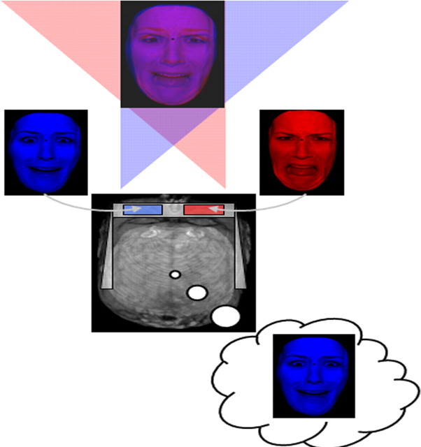
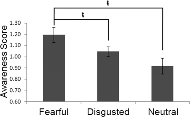
Emotional stimuli, including facial expressions, are thought to gain rapid and privileged access to processing resources in the brain. Despite this access, we are conscious of only a fraction of the myriad of emotion-related cues we face everyday. It remains unclear, therefore, what the relationship is between activity in neural regions associated with emotional representation and the phenomenological experience of emotional awareness. We used functional magnetic resonance imaging and binocular rivalry to delineate the neural correlates of awareness of conflicting emotional expressions in humans. Behaviorally, fearful faces were significantly more likely to be perceived than disgusted or neutral faces. Functionally, increased activity was observed in regions associated with facial expression processing, including the amygdala and fusiform gyrus during emotional awareness. In contrast, awareness of neutral faces and suppression of fearful faces were associated with increased activity in dorsolateral prefrontal and inferior parietal cortices. The amygdala showed increased functional connectivity with ventral visual system regions during fear awareness and increased connectivity with perigenual prefrontal cortex (pgPFC; Brodmann's area 32/10) when fear was suppressed. Despite being prioritized for awareness, emotional items were associated with reduced activity in areas considered critical for consciousness. Contributions to consciousness from bottom-up and top-down neural regions may be additive, such that increased activity in specialized regions within the extended ventral visual system may reduce demands on a frontoparietal system important for awareness. The possibility is raised that interactions between pgPFC and the amygdala, previously implicated in extinction, may also influence whether or not an emotional stimulus is accessible to consciousness.
Figures
 = p < 0.05).
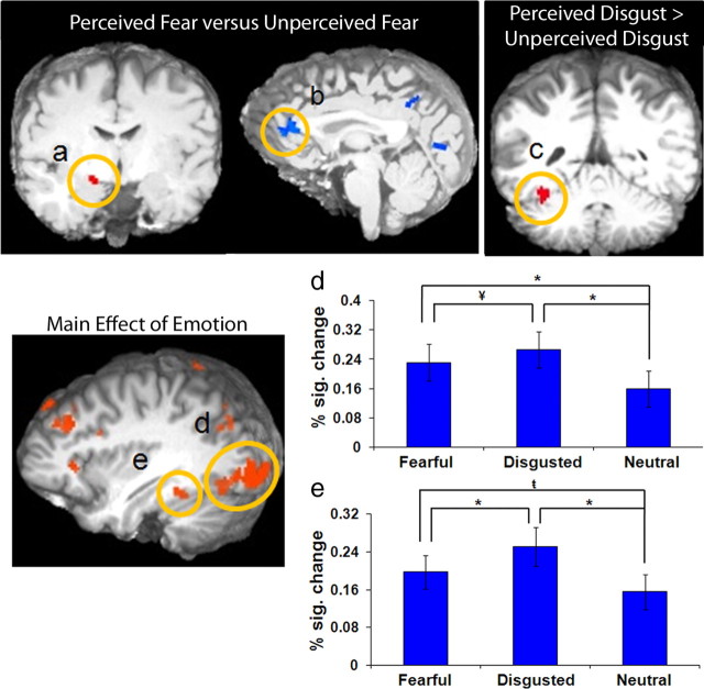 p < 0.05).
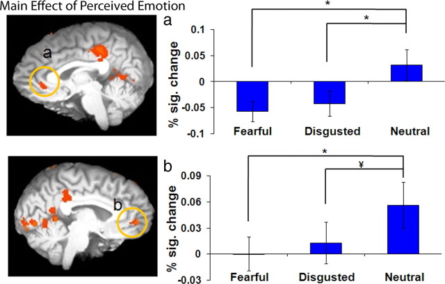
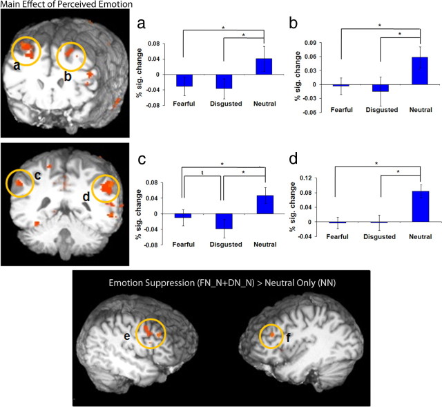 p < 0.05). e, f, The results of the contrast of emotion suppression [FN_N + DN_N] > neutral only condition (NN) revealed greater activation in the right (e) middle frontal gyrus and the left (f) middle frontal gyrus during emotion suppression. The y-axis depicts percentage signal change in BOLD response; error bars depict the SEM between subjects.
References
-
- Adolphs R, Tranel D, Damasio H, Damasio A. Impaired recognition of emotion in facial expressions following bilateral damage to the human amygdala. Nature. 1994;372:669–672. - PubMed
-
- Amting JM, Miller J, Chow M, Mitchell DGV. Getting mixed messages: the impact of conflicting social signals on the brain's target emotional response. Neuroimage. 2009;47:1950–1959. - PubMed
-
- Anderson AK, Phelps EA. Lesions of the human amygdala impair enhanced perception of emotionally salient events. Nature. 2001;411:305–309. - PubMed
-
- Bar M, Tootell RB, Schacter DL, Greve DN, Fischl B, Mendola JD, Rosen BR, Dale AM. Cortical mechanisms specific to explicit visual object recognition. Neuron. 2001;29:529–535. - PubMed
Publication types
MeSH terms
Substances
LinkOut - more resources
Full Text Sources