. 2010 Jul 28;30(30):10086-95.
doi: 10.1523/JNEUROSCI.1227-10.2010.

Chronic systemic infection exacerbates ischemic brain damage via a CCL5 (regulated on activation, normal T-cell expressed and secreted)-mediated proinflammatory response in mice

Affiliations

Chronic systemic infection exacerbates ischemic brain damage via a CCL5 (regulated on activation, normal T-cell expressed and secreted)-mediated proinflammatory response in mice

Adám Dénes et al. J Neurosci. .

Abstract

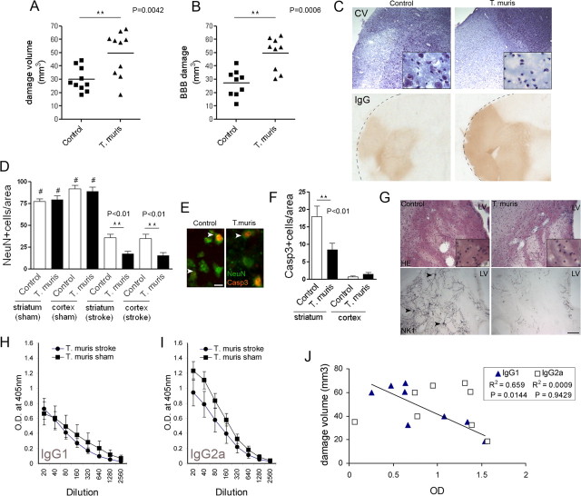
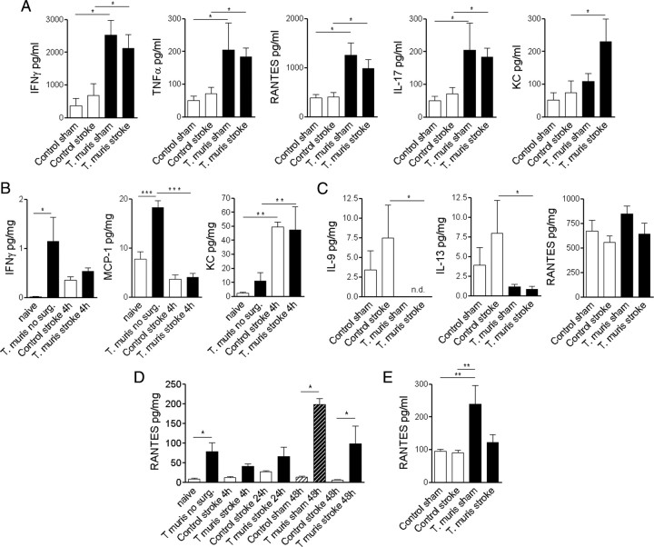
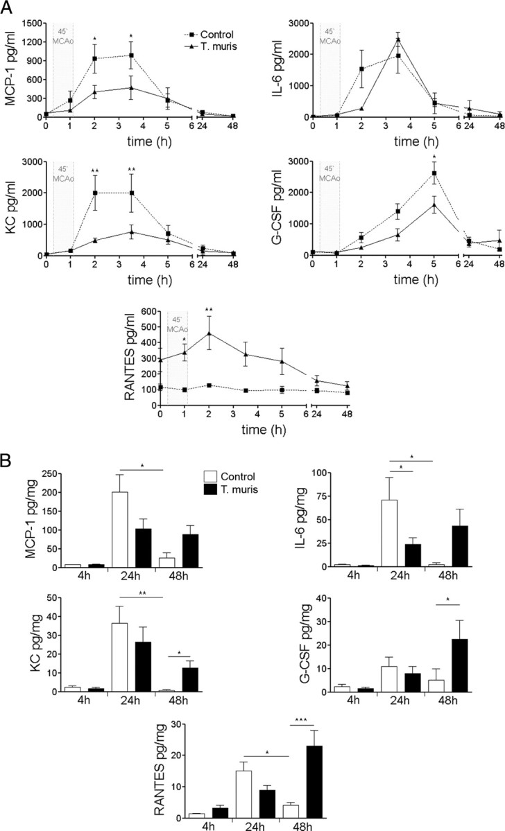
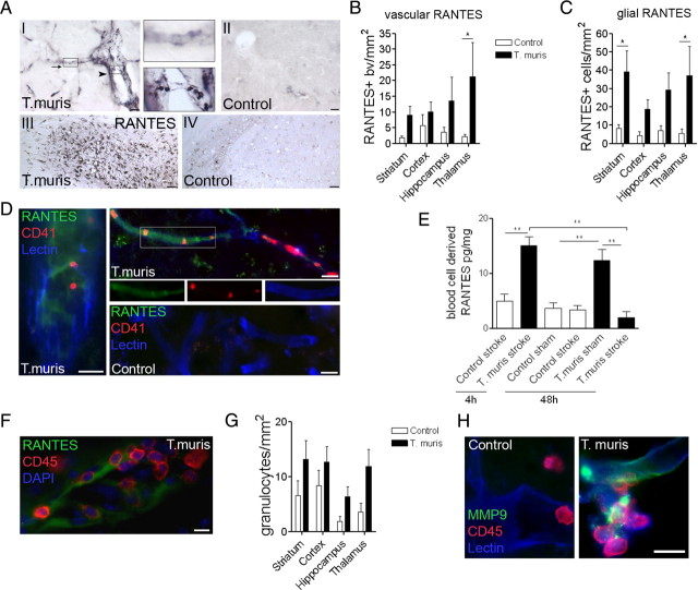
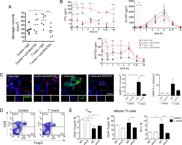
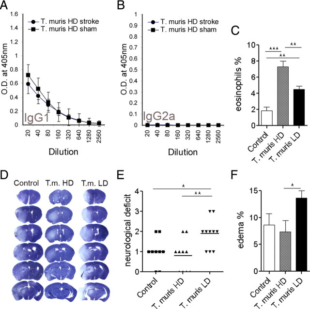
Infection and systemic inflammation are risk factors for cerebrovascular diseases and poststroke infections impair outcome in stroke patients, although the mechanisms of their contribution are mostly unknown. No preclinical studies have identified how chronic infection affects ischemic brain damage and which key inflammatory mediators are involved. We used a well established model of gut infection (Trichuris muris) to study how chronic infection contributes to brain injury. We show that, in mice, infection that leads to a chronic Th1-polarized immune response dramatically (60%) exacerbates brain damage caused by experimental stroke. Chronic Th1-type infection resulted in systemic upregulation of proinflammatory mediators and profoundly altered stroke-induced early (40 min to 4 h) and late (48 h) inflammation in the brain and peripheral tissues. Using the same infection, we show that a Th1-, but not Th2-polarized response augments brain injury by increasing the Th1 chemokine CCL5 [regulated on activation, normal T-cell expressed and secreted (RANTES)] systemically. This infection-associated response paralleled altered regulatory T-cell response, accelerated platelet aggregation in brain capillaries, and increased microvascular injury and matrix metalloproteinase activation after stroke. Antibody neutralization of RANTES reversed the effect of chronic infection on brain damage, microvascular MMP-9 activation, and cellular inflammatory response. Our results suggest that chronic infection exacerbates ischemic brain damage via a RANTES-mediated systemic inflammatory response, which leads to delayed resolution of inflammation and augmented microvascular injury in the brain.


Figures

Figure 1.
Figure 1.
Chronic Th1-type peripheral immune response augments ischemic brain damage. Ischemic (A) and BBB (B) damage are significantly larger in infected mice compared with uninfected mice at 48 h reperfusion after 45 min MCAo, as identified by cresyl violet staining (C) and leakage of plasma-derived IgG (t test, two-tailed). Quantitative analysis of NeuN (D) and cleaved caspase-3 (F) immunofluorescence (E) shows augmented neuronal loss after chronic infection (ANOVA, followed by Bonferroni's multiple-comparison test; #p < 0.001 sham vs stroke; **p < 0.01). G, Hematoxylin and eosin staining shows significant loss of neurons in the ipsilateral striatum at 48 h reperfusion after 45 min MCAo, which is more pronounced in infected mice. Chronic peripheral infection causes marked degeneration and loss of NK1-positive cell bodies (arrowheads) and neuronal processes after MCAo. Measurement of parasite-specific IgG1 (H) and IgG2a (I) indicates the development of a Th1-polarized response. J, Correlation analysis indicates significant negative correlation between anti-parasitic IgG1 (dominant in Th2-type response) levels in the plasma and the volume of ischemic damage. Data are expressed as means ± SEM. LV, Lateral ventricle. Scale bars: E, 10 μm; G, 100 μm. Data shown are representative of 10 mice in each group corresponding to two independent experiments.
Figure 2.
Figure 2.
Chronic Th1-type infection results in organ-specific proinflammatory activation and systemic upregulation of RANTES. A, Mesenteric lymph node cells isolated 48 h after sham surgery or MCAo were restimulated with parasite ES antigens in vitro. CBA analysis was performed on spleen homogenates of mice without surgery (0 h), at 4 h reperfusion (B), and after 48 h reperfusion (C). D, Liver homogenates harvested at various time points show chronically elevated RANTES in infected mice. E, Plasma samples show elevated RANTES in sham mice at 48 h reperfusion. *p < 0.05, **p < 0.01, ***p < 0.001, one-way ANOVA followed by Bonferroni's multiple-comparison test. Data are expressed as means ± SEM. Data shown are representative of 6–10 mice in each group from two independent experiments.
Figure 3.
Figure 3.
Chronic infection alters MCAo-induced acute phase response in the plasma and the brain. A, Serial plasma samples. B, Ipsilateral brain hemisphere homogenates. *p < 0.05, **p < 0.01, two-way ANOVA followed by Bonferroni's posttest. Data are expressed as means ± SEM. Data shown are representative of two independent experiments.
Figure 4.
Figure 4.
Chronic Th1-polarized response increases RANTES, platelet aggregation, leukocyte recruitment, and matrix metalloproteinase activity in the ischemic hemisphere. A, Immunohistochemistry reveals increased RANTES in microvessels (top inset, arrow) and platelets (bottom inset, arrowhead) in Th1-polarized mice (I), but not in uninfected animals (II) after MCAo. Glial RANTES expression was augmented in the affected hippocampus (III) of infected but not in uninfected mice (IV). Quantification of vascular (B) and glial (C) RANTES staining 48 h after MCAo (infected vs uninfected: p < 0.001, two-way ANOVA; Bonferroni's posttest, *p < 0.05). D, Vascular deposition of RANTES (green) and platelet aggregates (CD41, red) are increased in infected mice. E, Blood cell lysates contain elevated RANTES at 4 h reperfusion in infected mice compared with uninfected animals, which is reduced by 48 h reperfusion (**p < 0.01, one-way ANOVA followed by Bonferroni's multiple-comparison test). F, Vascular RANTES deposition was associated with leukocyte recruitment (CD45; red). G, Granulocyte numbers (SJC) were increased in the ipsilateral hemisphere in infected mice (infected vs control: p < 0.01, two-way ANOVA). H, Vascular MMP-9 (green) expression was enhanced after MCAo in the ischemic striatum of Th1-polarized mice. Scale bars: D, F, H, 10 μm; A, I, II, 20 μm; A, III, IV, 100 μm. Data are expressed as means ± SEM. Data shown are representative of 6–10 mice in each group from two independent experiments.
Figure 5.
Figure 5.
Systemic neutralization of RANTES reverses the effects of Th1-type infection on brain damage. A, The exacerbation of the ischemic brain damage is prevented by RANTES inhibition. *p < 0.05, **p < 0.01, one-way ANOVA followed by Bonferroni's multiple-comparison test. B, Effect of RANTES neutralization on plasma cytokine levels. Two-way ANOVA followed by Bonferroni's posttest. Infection versus control: p < 0.01 for IFN-γ, G-CSF, RANTES, two-way ANOVA (T. muris anti-RANTES vs *control anti-RANTES, #control IgG, T. muris IgG). C, Vascular MMP-9 immunostaining, expressed as the number of MMP-9-positive blood vessels (bv) per square millimeter, was significantly increased in infected, control IgG-treated mice in the ipsilateral hemisphere after MCAo, compared with uninfected animals. This was reversed in response to anti-RANTES treatment. The number of CD45/MMP-9-positive cells was not significantly different (right). Fluorescent micrographs showing MMP-9, CD45, and lectin triple-immunofluorescent staining in the ipsilateral striatum (left). D, Flow cytometric analysis shows that Th1-polarized mice have reduced CD4+/Foxp3+ (Treg) cells in the spleen 48 h after MCAo. E, The effect of systemic neutralization of RANTES on effector Th cells, Treg, and Gr1-positive leukocytes in the spleen. Scale bar, 50 μm. Data are expressed as means ± SEM. Data shown are representative of two independent experiments.
Figure 6.
Figure 6.
T. muris high-dose infection results in Th2-polarized immune response, which does not exacerbate brain damage. A, B, Antiparasitic IgG levels in the plasma of T. muris high-dose (HD)-infected (Th2-polarized) mice. C, Eosinophil granulocytes are elevated in the bone marrow in T. muris HD mice compared with uninfected (control) and T. muris low-dose (LD)-infected (Th1-polarized) mice. D, Representative cresyl violet-stained brain sections from uninfected, T. muris HD, and T. muris LD mice after 45 min MCAo and 48 h reperfusion. E, Neurological deficit is significantly exacerbated in T. muris LD mice compared with uninfected and T. muris HD mice. F, Brain edema is larger in T. muris LD mice compared with T. muris HD animals. *p < 0.05, **p < 0.01, ***p < 0.001, one-way ANOVA followed by Bonferroni's posttest. Data are expressed as means ± SEM. Data shown are representative of two independent experiments (n = 10).

References

    1. Alessi MC, Juhan-Vague I. Metabolic syndrome, haemostasis and thrombosis. Thromb Haemost. 2008;99:995–1000. - PubMed
    1. Anthony D, Dempster R, Fearn S, Clements J, Wells G, Perry VH, Walker K. CXC chemokines generate age-related increases in neutrophil-mediated brain inflammation and blood-brain barrier breakdown. Curr Biol. 1998;8:923–926. - PubMed
    1. Bancroft AJ, Else KJ, Grencis RK. Low-level infection with Trichuris muris significantly affects the polarization of the CD4 response. Eur J Immunol. 1994;24:3113–3118. - PubMed
    1. Bancroft AJ, Else KJ, Humphreys NE, Grencis RK. The effect of challenge and trickle Trichuris muris infections on the polarisation of the immune response. Int J Parasitol. 2001;31:1627–1637. - PubMed
    1. Bederson JB, Pitts LH, Tsuji M, Nishimura MC, Davis RL, Bartkowski H. Rat middle cerebral artery occlusion: evaluation of the model and development of a neurologic examination. Stroke. 1986;17:472–476. - PubMed

Publication types

MeSH terms