. 2010 Nov 25;116(22):4665-74.
doi: 10.1182/blood-2010-04-278184. Epub 2010 Jul 28.

Endothelium-derived but not platelet-derived protein disulfide isomerase is required for thrombus formation in vivo

Affiliations

Endothelium-derived but not platelet-derived protein disulfide isomerase is required for thrombus formation in vivo

Reema Jasuja et al. Blood. .

Abstract

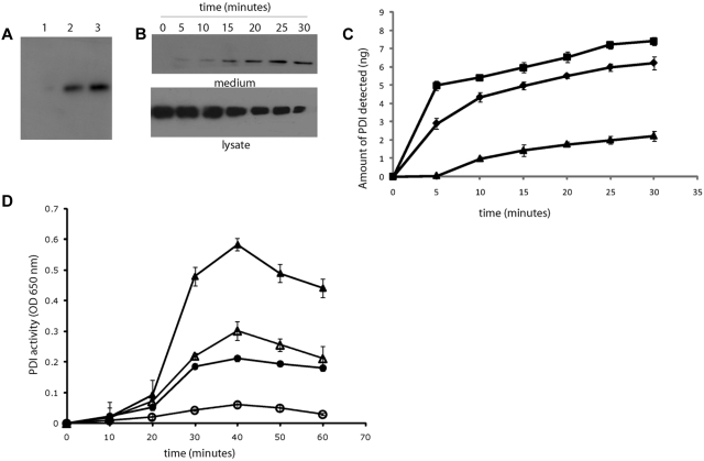
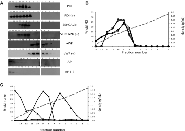
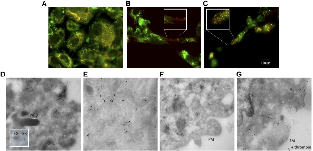
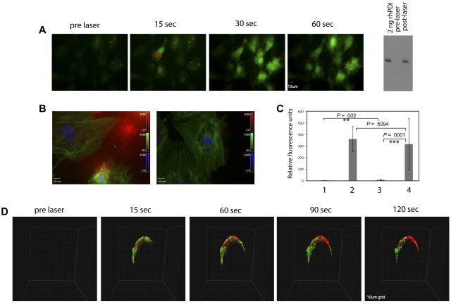
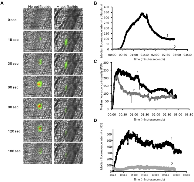
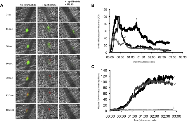
Protein disulfide isomerase (PDI) catalyzes the oxidation reduction and isomerization of disulfide bonds. We have previously identified an important role for extracellular PDI during thrombus formation in vivo. Here, we show that endothelial cells are a critical cellular source of secreted PDI, important for fibrin generation and platelet accumulation in vivo. Functional PDI is rapidly secreted from human umbilical vein endothelial cells in culture upon activation with thrombin or after laser-induced stimulation. PDI is localized in different cellular compartments in activated and quiescent endothelial cells, and is redistributed to the plasma membrane after cell activation. In vivo studies using intravital microscopy show that PDI appears rapidly after laser-induced vessel wall injury, before the appearance of the platelet thrombus. If platelet thrombus formation is inhibited by the infusion of eptifibatide into the circulation, PDI is detected after vessel wall injury, and fibrin deposition is normal. Treatment of mice with a function blocking anti-PDI antibody completely inhibits fibrin generation in eptifibatide-treated mice. These results indicate that, although both platelets and endothelial cells secrete PDI after laser-induced injury, PDI from endothelial cells is required for fibrin generation in vivo.


Figures

Figure 1
Figure 1
Agonist-induced PDI secretion from HUVECs. PDI secretion from HUVECs into serum-free cell culture medium after overnight culture of resting or activated cells was detected by sodium dodecyl sulfate–electrophoresis of the medium followed by immunoblotting with anti-PDI antibodies. (A) PDI in culture medium (50 μL from confluent monolayer ∼ 1 × 10⁶ cells) detected with the monoclonal anti-PDI antibody, RL90, at 1 μg/mL. Lane 1, conditioned media from resting HUVECs; lane 2, conditioned medium from HUVECs activated with 1 U/mL thrombin; lane 3, conditioned medium from HUVECs activated with 10μM A23187 calcium ionophore. (B) Immunoblot showing PDI secretion after thrombin stimulation from 5 × 10⁶ HUVECs over a period of 30 minutes. Top panel, medium from thrombin-activated HUVECs. Bottom panel, cell lysates from the corresponding activated cells. (C) Time course study of PDI release from 5 × 10⁶ HUVECs after stimulation with (■) 100 ng/mL PMA, (♦) 1 U/mL thrombin, or (▴) 0.1mM histamine. (D) PDI activity was measured by the insulin transhydrogenase assay in the conditioned media from 5 × 10⁶ thrombin-activated HUVECs (mean of 3 experiments) in the presence (unfilled) or absence (filled) of 5 μg/mL monoclonal inhibitory antibody RL90. Circles, PDI activity secreted from cultured HUVECs; triangles, activity of recombinant human PDI (1 μg/mL).
Figure 2
Figure 2
PDI distribution in resting and activated endothelial cells by gradient centrifugation. Organelles in a HUVEC lysate were separated by centrifugation on a Nycodenz density gradient. Percentages refer to percentage per fraction of total activity or antigen recovered in the 14 fractions of the gradient. The numbers 1-14 of the fractions are displayed on the x-axis. (A) Immunoblots of fractions 1-14 (higher to lower density, right to left) using the monoclonal PDI antibody, RL90, in fractions from unactivated (top panel, PDI) or thrombin-activated (second panel, PDI+) HUVECs. Middle and bottom panels show immunoblots for distribution of markers for endoplasmic reticulum, SERCA 2b; Weibel-Palade bodies, VWF; or plasma membrane, alkaline phosphatase, with (+) or without thrombin activation of cells. The numbers on the immunoblots refer to the density-gradient fractions. (B) The percentage per fraction of PDI antigen (—) or PDI enzymatic activity (- - -) are shown as the average of 2 experiments. The open circles (○) indicate fractions from unactivated HUVECs, while closed circles (•) show fractions from thrombin-activated HUVECs. The mean density of the fractions is indicated by the gray dashed line. (C). The average percentage per fraction from 2 experiments for markers, alkaline phosphatase (▴), SERCA2b (•), or VWF (■).
Figure 3
Figure 3
Cellular distribution of PDI in HUVECs. Intracellular localization of PDI was detected by immunostaining of fixed cultured HUVECs with monoclonal antibody RL90. (A) Simultaneous immunostaining of cells for PDI and SERCA2b in HUVECs indicates that these 2 proteins are colocalized in the endoplasmic reticulum. In addition, PDI is observed in granules distinct from the endoplasmic reticulum. Alexa 647–labeled anti-SERCA2b, red; Alexa 488–labeled RL90, green; colocalization, yellow. (B) Simultaneous immunostaining of cells for PDI and VWF indicates that PDI is not stored in Weibel Palade bodies. Alexa 647–labeled goat anti–rabbit IgG was used as a secondary antibody to detect VWF, red; Alexa 488–labeled RL90, green; colocalization, yellow. (C) Simultaneous immunostaining of chemokine Gro-α and PDI shows partial colocalization in small cytoplasmic granules. Original magnification in all panels ×60. Insets show high magnification (×100) of framed areas. (D) Immunogold labeling for PDI (10-nm gold particles) in HUVECs showed PDI in endoplasmic reticulum–related tubulovesicular structures and in small moderately electron-dense granules of approximately 100-150-nm diameter. WPB, Weibel-Palade bodies; ER, endoplasmic reticulum, SG, moderately electron-dense secretory granules (×99 000). (E) PDI and SERCA2b are colocalized in the endoplasmic reticulum, as indicated by immunogold staining. Only PDI is detected in secretory granules (×99 000). F) No plasma membrane–associated signal is detected in resting HUVECs (×119 000). (G) PDI, but not SERCA2b, is bound to the plasma membrane in activated HUVECs (×119 000). PM, plasma membrane. Panels E-G: PDI, 5-nm gold particles; SERCA2b, 15-nm gold particles.
Figure 4
Figure 4
Rapid calcium mobilization and PDI expression after laser injury in vitro and in vivo. Fluo-4-AM (3μM) was incubated with cultured HUVECs before laser injury and observed by fluorescence microscopy. (A) The images show a representative field of cells before and after a direct laser pulse to the point indicated by X. Cell imaging was initiated before laser injury to obtain a baseline image, and the laser was fired during this capture. Increased green signal in subsequent time-lapse images represents increased intracellular calcium monitored by Fluo-4 fluorescence, shown in representative images. The Western blot on the right depicts an immunoblot with polyclonal anti-PDI antibody to detect PDI in conditioned medium from unactivated (lane 2) or laser-activated (lane 3) HUVECs. Lane 1: 2 ng recombinant human PDI. (B) Representative images of fixed and immunostained cells that have been activated by laser injury in the presence of plasma and calcium with (right) or without (left) a function blocking PDI antibody RL90. The cells were fixed after laser activation and stained for fibrin (red). Fluorescein isothiocyanate–phalloidin (green) and DAPI (4,6 diamidino-2-phenylindole; blue). (C) Quantification of fibrin signal detected on cultured endothelial: lane 1, unactivated cells; lane 2, laser-activated cells; lane 3, laser-activated cells in the presence of the PDI-inhibitory antibody RL90; lane 4, laser-activated cells in the presence of an isotype control antibody. The P values between the different groups were obtained using the unpaired t test. (D) Representative images of rapid activation of arteriolar endothelium and PDI expression after laser injury in confocal intravital microscopy. Rhod-2 (6μM) and Alexa 647–labeled polyclonal anti-PDI antibody (0.3 μg/g body weight) were infused 5 minutes before the first injury. Site of injury is indicated by an X. Platelet aggregation was blocked with the GPIIbIIIa antagonist, eptifibatide. Vessel imaging was initiated before laser injury, and the laser was fired after one z-stack of 30 planes was obtained. Each z-stack of 30 images was 8.7 seconds. The grid size was 10 μm. Calcium elevation was monitored by excitation of Rhod-2 at 561 nm (pseudocolored green), while the PDI signal was observed simultaneously at 647 nm (pseudocolored red).
Figure 5
Figure 5
Comparison of PDI expression and platelet accumulation during thrombus formation. Rabbit polyclonal anti-PDI antibody conjugated to Alexa Fluor 488 (0.3 μg/g body weight) and Fab fragments of anti–CD-41 antibody conjugated to Alexa Fluor 647 (0.2 μg/g body weight) were infused into the mouse 5 minutes before arteriolar injury. In certain conditions, eptifibatide (10 μg/g body weight) was infused before injury and reinfused every 20 minutes for subsequent thrombi. (A). Representative binarized images of the appearance of fluorescence signals associated with PDI (green) and platelets (red) over 180 seconds after laser-induced vessel-wall injury in a wild-type (WT) mouse (left panels) or a wild-type mouse treated with eptifibatide. (B) Median integrated platelet fluorescence. Median fluorescence is presented vs. time after vessel-wall injury. Curve 1, WT mice; curve 2, WT mice treated with eptifibatide. (C-D) Median integrated PDI fluorescence detected by rabbit affinity-purified anti-PDI antibody. (D) Wild-type mouse treated with sodium beraprost (30 μg/kg body weight). Curve 1, WT mice; curve 2, WT mice treated with sodium beraprost.
Figure 6
Figure 6
Inhibition of fibrin formation with a function-blocking PDI antibody is platelet independent. Rabbit polyclonal anti-PDI antibody conjugated to Alexa Fluor 488 (0.3 μg/g body weight) and fibrin-specific mouse anti–human fibrin II β-chain monoclonal antibody conjugated to Alexa Fluor 647 (0.5 μg/g body weight) were infused into the mouse 5 minutes before arteriolar injury. (A) Representative binarized images of the appearance of fluorescence associated with PDI (green) or fibrin (red) are shown over 180 seconds after laser-induced vessel-wall injury in a wild-type mouse (left panel), a wild-type mouse treated with eptifibatide (10 μg/g body weight; middle panel), or a wild-type mouse treated with eptifibatide (10 μg/g body weight) and a function blocking anti-PDI antibody, RL90, (2 μg/g body weight; right panel). Inhibitory monoclonal anti-PDI antibody RL90 and/or eptifibatide were infused into the circulation 5 minutes before injury. Data were collected from the same mouse pre- and postinfusion of eptifibatide and/or RL90. (B) Median integrated PDI fluorescence intensity or (C) median integrated fibrin fluorescence intensity for thrombi formed before (curve 1) or after the infusion of eptifibatide (curve 2) or after the infusion of eptifibatide in the presence of RL90 (curve 3). Median fluorescence is presented versus time after vessel wall injury.

References

    1. Chen VM, Hogg PJ. Allosteric disulfide bonds in thrombosis and thrombolysis. J Thromb Haemost. 2006;4:2533–2541. - PubMed
    1. Chen C, Lin Y, Detwiler DC. Protein disulfide isomerase activity is released by activated platelets. Blood. 1992;79(9):2226–2228. - PubMed
    1. Essex DW, Chen K, Swiatkowska M. Localization of protein disulfide isomerase to the external surface of the platelet plasma membrane. Blood. 1995;86(6):2168–2173. - PubMed
    1. Burgess JK, Hotchkiss KA, Suter C, et al. Physical proximity and functional association of glyco-protein-1b alpha and protein disulfide isomerase on the platelet plasma membrane. J Biol Chem. 2000;275(13):9758–9766. - PubMed
    1. Hotchkiss KA, Matthias LJ, Hogg PJ. Exposure of the cryptic Arg-Gly-Asp sequence in thrombospondin-1 by protein disulfide isomerase. Biochim Biophys Acta. 1998;1388(2):478–488. - PubMed

Publication types