. 2010 Nov 11;116(19):3828-38.
doi: 10.1182/blood-2010-06-288068. Epub 2010 Jul 28.

Improved cellular and humoral immune responses in vivo following targeting of HIV Gag to dendritic cells within human anti-human DEC205 monoclonal antibody

Affiliations

Improved cellular and humoral immune responses in vivo following targeting of HIV Gag to dendritic cells within human anti-human DEC205 monoclonal antibody

Cheolho Cheong et al. Blood. .

Abstract

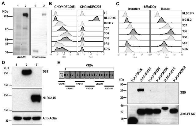
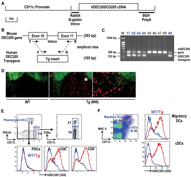
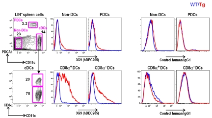
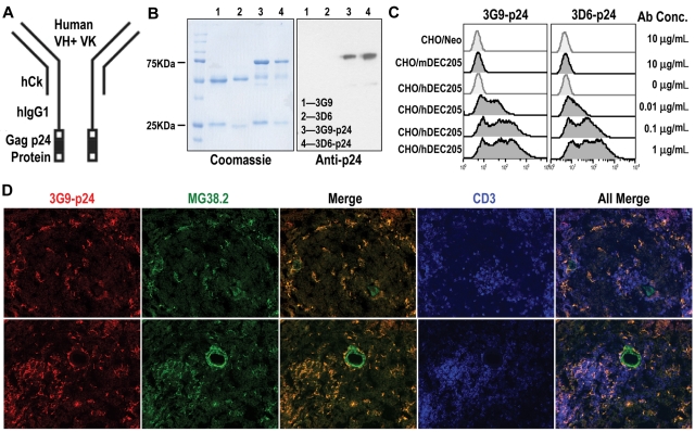
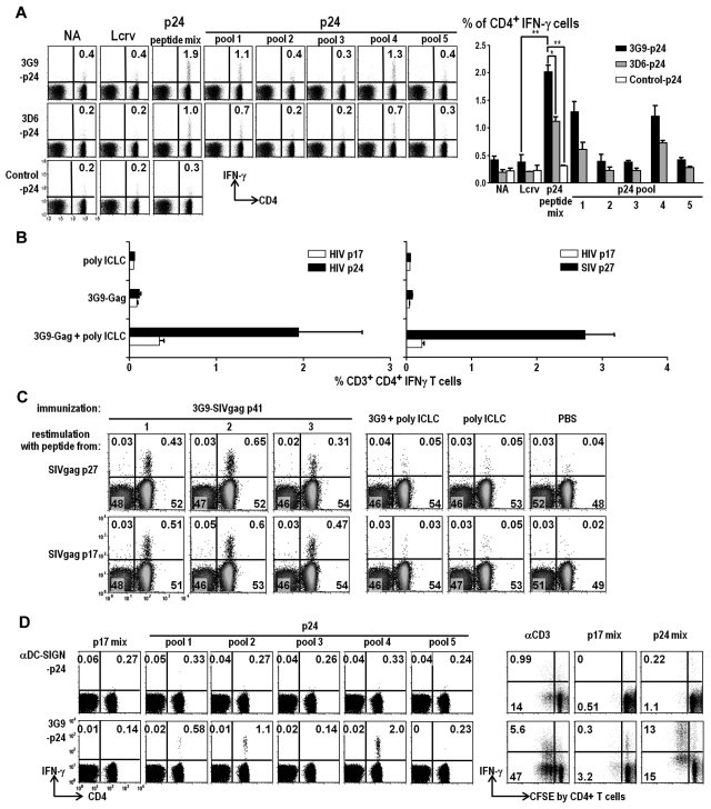
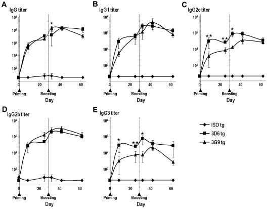
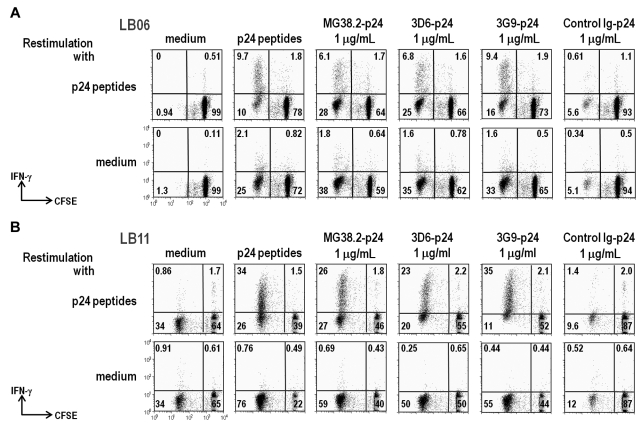
Protein vaccines for T-cell immunity are not being prioritized because of poor immunogenicity. To overcome this hurdle, proteins are being targeted to maturing dendritic cells (DCs) within monoclonal antibodies (mAbs) to DC receptors. To extend the concept to humans, we immunized human immunoglobulin-expressing mice with human DEC205 (hDEC205) extracellular domain. 3D6 and 3G9 mAbs were selected for high-affinity binding to hDEC205. In addition, CD11c promoter hDEC205 transgenic mice were generated, and 3G9 was selectively targeted to DCs in these animals. When mAb heavy chain was engineered to express HIV Gag p24, the fusion mAb induced interferon-γ- and interleukin-2-producing CD4(+) T cells in hDEC205 transgenic mice, if polynocinic polycytidylic acid was coadministered as an adjuvant. The T-cell response was broad, recognizing at least 3 Gag peptides, and high titers of anti-human immunoglobulin G antibody were made. Anti-hDEC205 also improved the cross-presentation of Gag to primed CD8(+) T cells from HIV-infected individuals. In all tests, 3D6 and 3G9 targeting greatly enhanced immunization relative to nonbinding control mAb. These results provide preclinical evidence that in vivo hDEC205 targeting increases the efficiency with which proteins elicit specific immunity, setting the stage for proof-of-concept studies of these new protein vaccines in human subjects.


Figures

Figure 1
Figure 1
Generation of human anti-hDEC205 monoclonal antibodies. (A) Expression of soluble human DEC205-human IgG1 Fc (V5.hDEC205/hIgG1Fc) fusion protein. Left: CHO cells, transfected with control plasmid (lane 1) and V5.hDEC205/hIgG1Fc (lane 2), were lysed and analyzed by Western blotting with anti-V5 antibody. Right: purified human IgG1 Fc alone (lane 1) or V5.hDEC205/hIgG1Fc fusion proteins (lane 2) produced by transfected CHO cells were analyzed on SDS-PAGE and stained by Coomassie blue. (B) FACS labeling of CHO/hDEC205 and CHO/mDEC205 cells and (C) human MoDCs stained at 1 μg/mL with human anti-hDEC205 mAbs, as well as the previously described NLDC145 rat anti-mDEC205 and MG38.2 mouse anti-hDEC205. The MoDCs were treated with/without LPS (100 ng/mL) to generate mature and immature DCs. (D) Lysates from CHO/Neo (lane 1), CHO/hDEC205 (lane 2), and CHO/mDEC205 (lane 3) cells were analyzed by Western blotting with mAbs 3G9 anti-hDEC205, NLDC145 anti-mDEC205, and anti-actin. (E) A series of deletion constructs in the extracellular domain of human DEC205 were generated (left, underlines), each with a FLAG tag, and each FLAG-tagged deletion construct was expressed in 293T cells. To map the 3G9 epitope on DEC205, each cell lysate was analyzed by Western blotting with mAbs 3G9 and L5 anti-FLAG.
Figure 2
Figure 2
Generation of hDEC205 transgenic mice. (A) Schematic of a linearized CD11c promoter-hDEC205 transgene. (B) PCR genotyping strategy for hDEC205 Tg mice, where a set of primers was selected for conserved sequences between mouse and human DEC205. The shorter (165 bp) PCR amplicon is generated from the hDEC205 intronless transgene and the longer (293 bp) from the mouse DEC205 gene. Arrows indicate the positions of F (forward) and R (reverse) primers. (C) A representative gel of 5 hDEC205 Tg founders after PCR of genomic DNA from newborn Tg mouse tails. M is for a lane loaded with molecular weight markers, and bold numbers (17, 42, 43, 44, and 49) indicate the lanes with samples from founder mice carrying the transgene. (D) Localization of hDEC205 in lymph nodes of hDEC205 Tg no. 49 and wild-type mice was visualized by immunohistochemistry. Acetone-fixed cryostat sections of subcutaneous lymph nodes were stained with 3G9 mAb, followed by secondary PE-conjugated antihuman IgG, and FITC-anti-B220 to discriminate B and T areas. Images were taken 200× magnification, but the region with the asterisk (*) is magnified 400× on the right using a Molecular Devices OlympusAX70 deconvolution microscope (Olympus) running METAMORPH Meta Imaging 3.0 (Universal Imaging Corporaration). (E) Expression of hDEC205 in the spleen of wild-type and hDEC205 Tg no. 49 mice. Lymphocyte lineage (CD3, CD19, and DX5) negative cells were surface stained with antibodies for hDEC205 (3G9-Alexa647), CD11c, CD8α, and PDCA1 to monitor hDEC205 expression in PDCs (PDCA1+), and cDCs (CD11c+ and CD8+ or CD8). (F) Skin-draining lymph node cell suspension from wild-type and hDEC205 Tg no. 49 mice were stained with mAbs for hDEC205 (3G9-A647), CD11c, and MHC II to detect hDEC205 in migratory DCs and cDCs. In histograms, blue is for wild-type (WT) and red for hDEC205 Tg mice (Tg; E-F).
Figure 3
Figure 3
Targeting of 3G9 to DCs in vivo. Wild-type and hDEC205 Tg mice were injected intraperitoneally with Alexa 647–labeled 3G9 or human IgG1 isotype (30 μg per mouse). Twelve hours later, lymphocyte lineage-negative (LIN) splenic cell suspensions were stained with the indicated antibodies. In the histograms, blue is for wild-type (WT) and red for hDEC205 Tg mice (Tg).
Figure 4
Figure 4
Expression and binding of hybrid anti-hDEC205-HIV Gag p24 fusion mAbs. (A) Schematic view of engineered mAb fused with HIV Gag p24. (B) Purified anti-hDEC205 mAbs 3D6 and 3G9 and their p24-fused hybrid mAbs were separated on 10% SDS-PAGE and stained with Coomassie blue (left) and blotted with anti-HIV Gag p24 antibodies (right). (C) Binding of hybrid mAbs to CHO/hDEC205, CHO/mDEC205, and control CHO/Neo cells using graded doses of purified recombinant p24-fused mAbs, followed by anti-hIgG-PE. (D) Detection of hDEC205 in normal human spleen sections after costaining with biotinylated 3G9-p24 (Alexa 555, red), MG38.2 mouse anti-hDEC205 (Alexa 488, green), and anti-hCD3 (APC, blue) to mark the T-cell areas. Images were taken 200× magnification using a Molecular Devices OlympusAX70 deconvolution microscope running METAMORPH Meta Imaging 3.0
Figure 5
Figure 5
hDEC205 targeting with adjuvant induces Gag-specific CD4+ T-cell responses. (A) hDEC205 Tg mice were injected intraperitonally with 10 μg of purified 3G9-HIV Gag p24, 3D6-p24, or control Ig-p24 fusion mAbs along with 50 μg of poly IC and 25 μg of anti-CD40 mAb. After 2 weeks, splenic T-cell responses to pools of p24 peptides were analyzed. FACS plots from a representative experiment and histograms from 3 independent experiments are shown in percentiles of CD3+, IFN-γ–producing CD4+ T cells; similar results were obtained for IL-2–producing cells (not shown). Asterisks for statistically significant changes (*P ≤ .05, **P ≤ .01). (B) As in panel A, but prime-boost immunizations with 3G9-HIV Gag p24 (left, in blue) or 3G9-SIV Gag p27 (right, in red), plus only poly ICLC as an adjuvant. (C) SIV Gag p41 was the immunizing antigen to test responses to p17 and p27 regions of Gag. (D) As in panel A, showing IFN-γ–producing T cells after immunization with 3G9-HIV Gag p24, but not control anti-human DC-SIGN-p24 (left), and also specific proliferation (CFSE dilution) to HIV Gag p24, not HIV Gag p17 peptides (right). hDEC205 Tg in C57BL/6 background (A-C) and crossed with B10.BR (D).
Figure 6
Figure 6
Targeting to hDEC205 enhances humoral responses. At day 0, hDEC205 Tg mice were primed by intraperitonal injection with 10 μg of purified 3G9, 3D6, or control human IgG1 mAbs in the presence of 50 μg of poly IC and 25 μg of anti-CD40 mAb. At day 25, mice were boosted intraperitoneally with 10 μg of the same antibodies without adjuvants. Serum samples were collected at days −2, 5, 21, 31, 40, and 58. Serum antihuman IgG1 titers of total mouse IgG (A), IgG1 (B), IgG2c (C), IgG2b (D), and IgG3 (E) were measured by ELISA. Filled diamonds, squares, and triangles represent each group (3 mice per group). Asterisks denote statistical significances (*P ≤ .05; **P ≤ .01).
Figure 7
Figure 7
Improved cross-presentation of Gag protein via DEC205 to human T cells. (A) PBMCs from a long-term nonprogressor patient (LB06) were CFSE labeled and cultured 7 days with the indicated sources of HIV Gag p24 antigen (top). At the end of the culture, the cells were restimulated 6 hours with a pool of 50 Gag 15-mer peptides (upper) or without peptides (lower) in the presence of anti-CD28 costimulatory mAb. Data show IFN-γ production by CD8+ T cells, most of which have extensively diluted their CFSE label as a result of cell division. (B) As in (A), but the cells are mixtures of MoDCs and autologous T cells from a chronically HIV-infected donor (LB11). The data are displayed as 2 color dot plots to measure CFSE dilution and IFN-γ production in CD3+CD8+ cells.

References

    1. Hawiger D, Inaba K, Dorsett Y, et al. Dendritic cells induce peripheral T cell unresponsiveness under steady state conditions in vivo. J Exp Med. 2001;194(6):769–780. - PMC - PubMed
    1. Bonifaz L, Bonnyay D, Mahnke K, Rivera M, Nussenzweig MC, Steinman RM. Efficient targeting of protein antigen to the dendritic cell receptor DEC-205 in the steady state leads to antigen presentation on major histocompatibility complex class I products and peripheral CD8+ T cell tolerance. J Exp Med. 2002;196(12):1627–1638. - PMC - PubMed
    1. Bonifaz LC, Bonnyay DP, Charalambous A, et al. In vivo targeting of antigens to maturing dendritic cells via the DEC-205 receptor improves T cell vaccination. J Exp Med. 2004;199(6):815–824. - PMC - PubMed
    1. Caminschi I, Lahoud MH, Shortman K. Enhancing immune responses by targeting antigen to DC. Eur J Immunol. 2009;39(4):931–938. - PubMed
    1. Boscardin SB, Trumpfheller C, Nussenzweig MC, Steinman RM. Vaccines based on dendritic cell biology. In: Levine MM, editor. New Generation Vaccines. New York, NY: Informa Healthcare; 2009. pp. 327–339. Vol Chapter 32.

Publication types

MeSH terms