. 2010 Aug 17;107(33):14535-40.
doi: 10.1073/pnas.1004748107. Epub 2010 Jul 28.

Second harmonic generating (SHG) nanoprobes for in vivo imaging

Affiliations

Second harmonic generating (SHG) nanoprobes for in vivo imaging

Periklis Pantazis et al. Proc Natl Acad Sci U S A. .

Abstract

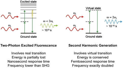
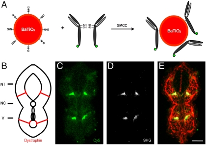
Fluorescence microscopy has profoundly changed cell and molecular biology studies by permitting tagged gene products to be followed as they function and interact. The ability of a fluorescent dye to absorb and emit light of different wavelengths allows it to generate startling contrast that, in the best cases, can permit single molecule detection and tracking. However, in many experimental settings, fluorescent probes fall short of their potential due to dye bleaching, dye signal saturation, and tissue autofluorescence. Here, we demonstrate that second harmonic generating (SHG) nanoprobes can be used for in vivo imaging, circumventing many of the limitations of classical fluorescence probes. Under intense illumination, such as at the focus of a laser-scanning microscope, these SHG nanocrystals convert two photons into one photon of half the wavelength; thus, when imaged by conventional two-photon microscopy, SHG nanoprobes appear to generate a signal with an inverse Stokes shift like a fluorescent dye, but with a narrower emission. Unlike commonly used fluorescent probes, SHG nanoprobes neither bleach nor blink, and the signal they generate does not saturate with increasing illumination intensity. The resulting contrast and detectability of SHG nanoprobes provide unique advantages for molecular imaging of living cells and tissues.


Conflict of interest statement

Conflict of interest statement: This work is the subject of patent applications filed by the California Institute of Technology.

Figures

Fig. 1.
Fig. 1.
Two-photon excited fluorescence versus SHG. Displayed are the Perrin–Jablonski fluorescence diagram (Left) and the energy-level diagram (Right) describing two-photon excited fluorescence and SHG, respectively. When intense light is shone on materials that do not possess an inversion symmetry, the vibrating electric field of the incident beam results in the polarization of the medium, reemitting light at the original frequency ωi but also at the frequency 2ωi (here shown, Right) that is twice the original one (with half of the wavelength). Unlike two-photon excited fluorescence, all of the incident radiation energy at frequency ωi is converted in the process of SHG to radiation at the SHG frequency 2ωi. And whereas two-photon excited fluorescence involves real energy transition of electrons, SHG involves only virtual energy transition. As a result, using ultrafast (femtosecond) pulsed lasers, the response time of SHG is at the femtosecond level, about several orders of magnitude faster than the nanosecond response time of fluorescence, allowing very fast and sensitive detection.
Fig. 2.
Fig. 2.
SHG nanoprobes signal properties. (A) Displayed is the normalized SHG signal spectrum of BaTiO3 nanocrystals of the size of around 30 nm (Inset, SEM picture of BaTiO3 nanocrystals) immobilized in 20% polyacrylamide gel (signal ranging from 380 to 710 nm) generated by tuning the excitation wavelength to 820 nm. (B) BaTiO3 nanocrystal immobilized in 20% polyacrylamide gel and illuminated with low-intensity levels of 820 nm light 300 times with a scanning speed of 20 frames/s. The SHG signal intensity of BaTiO3 is constant and does not display blinking. (C) BaTiO3 nanocrystals were immobilized in 20% polyacrylamide gel and rapidly illuminated with gradually increasing 820-nm light intensity. The SHG signal intensity of BaTiO3 nanocrystals increases quadratically. The dots show independent experimental results, whereas the line is a quadratic fit for BaTiO3 (n = 4). Bar shows direct comparison of the obtained intensity range of QDs (see E) when rapidly illuminated under comparable conditions with increasing (up to 40-fold) 820-nm light intensity. (D) Water-soluble CdSe/ZnS QD immobilized in 20% polyacrylamide gel and illuminated with low-intensity levels of 820-nm light 300 times with a scanning speed of 20 frames/s. In contrast to BaTiO3, the QD signal fluctuates displaying subblinking as well as major blinking. (E) QDs were immobilized in 20% polyacrylamide gel and rapidly illuminated with gradually increasing 820-nm light intensity. Signal saturation of the QD fluorescence occurs at very low-power levels. The dots show independent experimental results, whereas the line is a polynomial fit for QDs (n = 4).
Fig. 3.
Fig. 3.
SHG nanoprobes can be imaged with high SNR and virtually no signal background (AD). Using optimized emission filter sets SHG nanoprobes provide superior SNR in highly scattering and absorbing environments. BaTiO3 nanocrystals (A and A′) and water-soluble CdSe/ZnS quantum dots (B and B′) were immobilized in 2.5% Intralipid/0.05% Indian Ink/20% polyacrylamide gel, illuminated with 15% of 820-nm light, and respective signal intensity recorded either with a long (A and B) or a narrow (A′ and B′) bandpass emission filter. The SNR for BaTiO3 nanoparticles improves in this particular case 3-fold when a narrow emission filter is used (C) blocking most background signal from random absorption due to intense scattering, yet allowing transmission of a major part of SHG signal (A′, image). The SNR for QDs however cannot be increased when a narrow filter is used (D). In this case not only the background signal, but also the signal emission is significantly reduced (B′, image). Consequently, only BaTiO3 can be imaged with virtually no background signal while in parallel increasing the SNR. (AB′) Gray areas in the emission spectra of BaTiO3 (blue line) and QDs (red line) indicate blocked transmission by emission filters. (C and D) The SNR columns represent the means ± range of two measurements each. Bar = 500 nm.
Fig. 4.
Fig. 4.
SHG nanoprobes provide superior signal-to-noise ratio after in vivo injections BaTiO3 nanocrystals were injected into one-cell stage zebrafish embryos. Several days after cytoplasmic injection (around 72 hpf) excitation of nanocrystals with femtosecond pulsed 820-nm light results in strong SHG signal detectable in epidirection as well as in transdirection (A) throughout the whole zebrafish body [here, nanocrystals (arrowheads) present in the trunk of a zebrafish (BC′)]. Endogenous SHG from trunk muscles can only be detected in transdirection with the sarcomere repeat patterns clearly observable (B′). (D) BODIPY TR methyl ester dye labeling the extracellular matrix and cell membranes. (E) Merge of pictures B′, C′, and D. Note that the power levels required to detect endogenous SHG are 10 times higher than those to visualize BaTiO3. The SHG signal intensity of BaTiO3 is comparable in epi- as well as in transdetection mode. Anterior to the left. Bar = 75 μm.
Fig. 5.
Fig. 5.
SHG nanoprobe targeting specificity. (A) Scheme of SHG nanoprobe conjugation to Cy5-coupled (green pentagon) antibody fragments via disulphide reduction and sulfhydryl-amine coupling. (B) Schematic representation of a transversal section of trunk of a 24 hpf zebrafish embryo showing Dystrophin protein localization to fiber ends (red). NT, neural tube; NC, notochord; V, vessels. (CE) Immunostaining showing Dystrophin protein localization in a transversal tissue section using secondary antibody coupled to Cy5 (green) and BaTiO3 (white). Both readouts—Cy5 immunofluorescence (C), and BaTiO3 SHG signal (D)—label specifically Dystrophin. (E) Phalloidin labeling (red) is superimposed to show cell profiles in the transversal tissue section. Note that SHG immunostaining provides superior SNR of Dystrophin detection using a narrow emission filter. Bar = 30 μm.

Comment in

  • Microscope harmonies.
    Evanko D. Evanko D. Nat Methods. 2010 Oct;7(10):779. doi: 10.1038/nmeth1010-779. Nat Methods. 2010. PMID: 20936770 No abstract available.

References

    1. Prasher DC, Eckenrode VK, Ward WW, Prendergast FG, Cormier MJ. Primary structure of the Aequorea victoria green-fluorescent protein. Gene. 1992;111(2):229–233. - PubMed
    1. Shaner NC, Steinbach PA, Tsien RY. A guide to choosing fluorescent proteins. Nat Methods. 2005;2(12):905–909. - PubMed
    1. Lippincott-Schwartz J, Snapp E, Kenworthy A. Studying protein dynamics in living cells. Nat Rev Mol Cell Biol. 2001;2(6):444–456. - PubMed
    1. Hadjantonakis AK, Dickinson ME, Fraser SE, Papaioannou VE. Technicolour transgenics: Imaging tools for functional genomics in the mouse. Nat Rev Genet. 2003;4(8):613–625. - PubMed
    1. Baskin JM, et al. Copper-free click chemistry for dynamic in vivo imaging. Proc Natl Acad Sci USA. 2007;104(43):16793–16797. - PMC - PubMed

Publication types

MeSH terms