. 2010 Aug 17;75(7):611-8.
doi: 10.1212/WNL.0b013e3181ed9cde. Epub 2010 Jul 28.

Juvenile ALS with basophilic inclusions is a FUS proteinopathy with FUS mutations

Affiliations

Juvenile ALS with basophilic inclusions is a FUS proteinopathy with FUS mutations

D Bäumer et al. Neurology. .

Abstract

Background: Juvenile amyotrophic lateral sclerosis (ALS) with basophilic inclusions is a form of ALS characterized by protein deposits in motor neurons that are morphologically and tinctorially distinct from those of classic sporadic ALS. The nosologic position of this type of ALS in the molecular pathologic and genetic classification of ALS is unknown.

Methods: We identified neuropathologically 4 patients with juvenile ALS with basophilic inclusions and tested the hypothesis that specific RNA binding protein pathology may define this type of ALS. Immunohistochemical findings prompted us to sequence the fused in sarcoma (FUS) gene.

Results: Motor symptoms began between ages 17 and 22. Disease progression was rapid without dementia. No family history was identified. Basophilic inclusions were strongly positive for FUS protein but negative for TAR DNA binding protein 43 (TDP-43). Granular and compact FUS deposits were identified in glia and neuronal cytoplasm and nuclei. Ultrastructure of aggregates was in keeping with origin from fragmented rough endoplasmic reticulum. Sequencing of all 15 exons of the FUS gene in 3 patients revealed a novel deletion mutation (c.1554_1557delACAG) in 1 individual and the c.1574C>T (P525L) mutation in 2 others.

Conclusion: Juvenile ALS with basophilic inclusions is a FUS proteinopathy and should be classified as ALS-FUS. The FUS c.1574C>T (P525L) and c.1554_1557delACAG mutations are associated with this distinct phenotype. The molecular genetic relationship with frontotemporal lobar degeneration with FUS pathology remains to be clarified.


Figures

None
Figure 1 Neuropathology of amyotrophic lateral sclerosis (ALS) with basophilic inclusions and FUS mutations (A) No evidence of frontotemporal atrophy (case 1, P525L mutation). Severe (B, case 1) and mild (C, case 2) degeneration of the corticospinal tract in the P525L FUS mutation. (D–F) Compact basophilic neuronal cytoplasmic inclusions (arrows) were present in upper and lower motor neurons of all cases with FUS mutations. (D) Case 2, Betz cell, (E) case 4, Betz cell, (F) case 1, nucleus hypoglossus. (G) Fragments of straight filaments associated with electron-dense granules characterize a spinal cord neuronal cytoplasmic inclusion in case 1 with the FUS P525L mutation (×15,000 magnification). Luxol fast blue cresyl violet (B, C), hematoxylin-eosin (D–F).
None
Figure 2 Morphologic and immunohistochemical characterization of FUS pathology (A) A normal lower motor neuron from a control case. Note the thin crisp nuclear membrane and basophilic Nissl substance at the periphery of the cytoplasm. A consistent feature of FUS mutation cases on routine hematoxylin-eosin and cresyl violet stains was disruption, clumping, and redistribution of Nissl substance in motor neurons (arrows, B, C). p62 (sequestosome-1) antibody does not normally label Nissl substance but does so in a neuron in a FUS P525L case, which also shows a small aggregation focus (arrow, D). Numerous presumed pathologic FUS protein deposits can be seen when antibodies are titrated in a way that does not stain normal diffuse nuclear FUS (E–L). (E) Overview of the hypoglossal nucleus in FUS P525L case 1. (F–I) Granular (F) to increasingly compact cytoplasmic FUS deposition (G–H); P525L mutation. The latter correlates with the large basophilic inclusions that define this type of amyotrophic lateral sclerosis (I, inset). Nuclear (arrowheads, J) and glial (arrows, J) as well as neuritic (arrows, K) FUS pathology is also present. Note granular perinuclear neuronal FUS deposit (arrowhead, K). (L) Only case 3 had extensive neuronal and glial (arrow) FUS pathology outside the corticospinal system, and the substantia nigra is shown here.
None
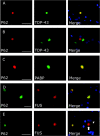
Figure 3 Characterization of FUS pathology by immunofluorescence (A) Compact cytoplasmic inclusions in classic sporadic amyotrophic lateral sclerosis are labeled with p62 and TDP-43. (B) Compact basophilic inclusions are also labeled by p62, but are negative for TDP-43, which retains its normal nuclear distribution. (C) Most basophilic inclusions are immunostained with PABP antibody, indicating the presence of polyadenylated RNA. (D) Basophilic inclusions that are p62 positive are also FUS positive (D), but not all FUS inclusions contain p62 (E, arrow). FUS nuclear staining is sometimes not completely abolished in cells with cytoplasmic FUS inclusions (E, arrowhead).
None
Figure 4 Genetics of juvenile amyotrophic lateral sclerosis (ALS) with basophilic inclusions (A) Sequence traces of control and patient DNA showing a 4–base pair deletion in FUS exon 15 (c.1554_1557delACAG) confirmed by sequence analysis of cloned wild-type (insert top, bases deleted in the mutant allele are marked by the box) and mutant allele (insert bottom). The deletion is predicted to cause a frameshift with resulting alteration of the terminal 8 amino acids of FUS (bottom). (B) Two patients were heterozygous for the c.1574C>T (P525L) mutation (top, control, bottom, case), altering the penultimate amino acid of FUS in an evolutionary conserved residue (C). The FUS protein structure according to reference sequence PRO_0000081591 (http://www.uniprot.org/uniprot/) is shown in (D). An asterisk marks residues previously found to be mutated in patients with familial or sporadic ALS. aa = amino acid.

Comment in

References

    1. Alonso A, Logroscino G, Jick SS, Hernan MA. Incidence and lifetime risk of motor neuron disease in the United Kingdom: a population-based study. Eur J Neurol 2009;16:745–751. - PMC - PubMed
    1. O'Toole O, Traynor BJ, Brennan P, et al. Epidemiology and clinical features of amyotrophic lateral sclerosis in Ireland between 1995 and 2004. J Neurol Neurosurg Psychiatry 2008;79:30–32. - PubMed
    1. Simpson CL, Al-Chalabi A. Amyotrophic lateral sclerosis as a complex genetic disease. Biochim Biophys Acta 2006;1762:973–985. - PubMed
    1. Rosen DR, Siddique T, Patterson D, et al. Mutations in Cu/Zn superoxide dismutase gene are associated with familial amyotrophic lateral sclerosis. Nature 1993;362:59–62. - PubMed
    1. Sreedharan J, Blair IP, Tripathi VB, et al. TDP-43 mutations in familial and sporadic amyotrophic lateral sclerosis. Science 2008;319:1668–1672. - PMC - PubMed

Publication types

MeSH terms

Substances