The effect of bilateral isometric forces in different directions on motor cortical function in humans
- PMID: 20668276
- PMCID: PMC3007641
- DOI: 10.1152/jn.00020.2010
The effect of bilateral isometric forces in different directions on motor cortical function in humans
Abstract
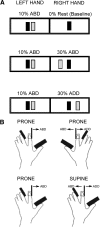
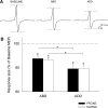
The activity in the primary motor cortex (M1) reflects the direction of movements, but little is known about physiological changes in the M1 during generation of bilateral isometric forces in different directions. Here, we used transcranial magnetic stimulation to examine motor evoked potentials (MEPs), short-interval intracortical inhibition (SICI), and interhemispheric inhibition (IHI) in the left first dorsal interosseous (FDI) during isometric index finger abduction while the right index finger remained at rest or performed isometric forces in different directions (abduction or adduction) and in different postures (prone and supine). Left FDI MEPs were suppressed during bilateral compared with unilateral forces, with a stronger suppression when the right index finger force was exerted in the adduction direction regardless of hand posture. IHI targeting the left FDI increased during bilateral compared with unilateral forces and this increase was stronger during right index finger adduction despite the posture of the right hand. SICI decreased to a similar extent during both bilateral forces in both hand postures. Thus generation of index finger isometric forces away from the body midline (adduction direction), regardless of the muscle engaged in the task, down-regulates corticospinal output in the contralateral active hand to a greater extent than forces exerted toward the body midline (abduction direction). Transcallosal inhibition, but not GABAergic intracortical circuits, was modulated by the direction of the force. These findings suggest that during generation of bimanual isometric forces the M1 is driven by "extrinsic" parameters related to the hand action.
Figures


Similar articles
-
Physiological changes underlying bilateral isometric arm voluntary contractions in healthy humans.J Neurophysiol. 2011 Apr;105(4):1594-602. doi: 10.1152/jn.00678.2010. Epub 2011 Jan 27. J Neurophysiol. 2011. PMID: 21273315 Free PMC article. Clinical Trial.
-
Progressive suppression of intracortical inhibition during graded isometric contraction of a hand muscle is not influenced by hand preference.Exp Brain Res. 2007 Feb;177(2):266-74. doi: 10.1007/s00221-006-0669-2. Epub 2006 Sep 1. Exp Brain Res. 2007. PMID: 16947062
-
Bimanual coordination of force enhances interhemispheric inhibition between the primary motor cortices.Neuroreport. 2014 Oct 22;25(15):1203-7. doi: 10.1097/WNR.0000000000000248. Neuroreport. 2014. PMID: 25144392
-
Mechanisms underlying functional changes in the primary motor cortex ipsilateral to an active hand.J Neurosci. 2008 May 28;28(22):5631-40. doi: 10.1523/JNEUROSCI.0093-08.2008. J Neurosci. 2008. PMID: 18509024 Free PMC article.
-
Organization of ipsilateral excitatory and inhibitory pathways in the human motor cortex.J Neurophysiol. 2003 Mar;89(3):1256-64. doi: 10.1152/jn.00950.2002. Epub 2002 Oct 30. J Neurophysiol. 2003. PMID: 12611955
Cited by
-
The effect of motor overflow on bimanual asymmetric force coordination.Exp Brain Res. 2017 Apr;235(4):1097-1105. doi: 10.1007/s00221-016-4867-2. Epub 2017 Jan 16. Exp Brain Res. 2017. PMID: 28091708 Free PMC article.
-
Corticomuscular coherence during bilateral isometric arm voluntary activity in healthy humans.J Neurophysiol. 2012 Apr;107(8):2154-62. doi: 10.1152/jn.00722.2011. Epub 2012 Jan 25. J Neurophysiol. 2012. PMID: 22279195 Free PMC article.
-
Bilateral deficit in maximal force production.Eur J Appl Physiol. 2016 Dec;116(11-12):2057-2084. doi: 10.1007/s00421-016-3458-z. Epub 2016 Aug 31. Eur J Appl Physiol. 2016. PMID: 27582260 Review.
-
Bilateral reach-to-grasp movement asymmetries after human spinal cord injury.J Neurophysiol. 2016 Jan 1;115(1):157-67. doi: 10.1152/jn.00692.2015. Epub 2015 Oct 14. J Neurophysiol. 2016. PMID: 26467518 Free PMC article.
-
Two distinct ipsilateral cortical representations for individuated finger movements.Cereb Cortex. 2013 Jun;23(6):1362-77. doi: 10.1093/cercor/bhs120. Epub 2012 May 17. Cereb Cortex. 2013. PMID: 22610393 Free PMC article.
References
-
- Ashe J. Force and the motor cortex. Behav Brain Res 87: 255–269, 1997 - PubMed
-
- Bäumer T, Dammann E, Bock F, Klöppel S, Siebner HR, Münchau A. Laterality of interhemispheric inhibition depends on handedness. Exp Brain Res 180: 195–203, 2007 - PubMed
-
- Cardoso de Oliveira S, Gribova A, Donchin O, Bergman H, Vaadia E. Neural interactions between motor cortical hemispheres during bimanual and unimanual arm movements. Eur J Neurosci 14: 1881–1896, 2001 - PubMed
-
- Carson RG. The dynamics of isometric bimanual coordination. Exp Brain Res 105: 465–476, 1995 - PubMed
-
- Carson RG. Neural pathways mediating bilateral interactions between the upper limbs. Brain Res Brain Res Rev 49: 641–662, 2005 - PubMed
Publication types
MeSH terms
Grants and funding
LinkOut - more resources
Full Text Sources

