Review
. 2011 Jan;36(1):52-73.
doi: 10.1038/npp.2010.104. Epub 2010 Jul 28.

Modes and models of forebrain cholinergic neuromodulation of cognition

Affiliations
Review

Modes and models of forebrain cholinergic neuromodulation of cognition

Michael E Hasselmo et al. Neuropsychopharmacology. 2011 Jan.

Abstract

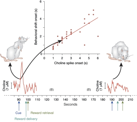
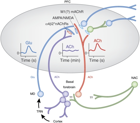
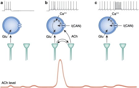
As indicated by the profound cognitive impairments caused by cholinergic receptor antagonists, cholinergic neurotransmission has a vital role in cognitive function, specifically attention and memory encoding. Abnormally regulated cholinergic neurotransmission has been hypothesized to contribute to the cognitive symptoms of neuropsychiatric disorders. Loss of cholinergic neurons enhances the severity of the symptoms of dementia. Cholinergic receptor agonists and acetylcholinesterase inhibitors have been investigated for the treatment of cognitive dysfunction. Evidence from experiments using new techniques for measuring rapid changes in cholinergic neurotransmission provides a novel perspective on the cholinergic regulation of cognitive processes. This evidence indicates that changes in cholinergic modulation on a timescale of seconds is triggered by sensory input cues and serves to facilitate cue detection and attentional performance. Furthermore, the evidence indicates cholinergic induction of evoked intrinsic, persistent spiking mechanisms for active maintenance of sensory input, and planned responses. Models have been developed to describe the neuronal mechanisms underlying the transient modulation of cortical target circuits by cholinergic activity. These models postulate specific locations and roles of nicotinic and muscarinic acetylcholine receptors and that cholinergic neurotransmission is controlled in part by (cortical) target circuits. The available evidence and these models point to new principles governing the development of the next generation of cholinergic treatments for cognitive disorders.


Figures

Figure 1
Figure 1
Prefrontal cholinergic transients mediating the detection of cues (data and components of this figure were adopted from Parikh et al, 2007). The abscissa depicts the time (seconds) over two trials, one in which the cue was detected (left) and one in which the cue was missed (right). Animals performed a cued appetitive response task. A light cue (presented for 1 s; blue arrows) predicted reward delivery 6±2 s later at one out of two reward ports (dark green arrows). Detection was defined behaviorally by cue-evoked orientation toward and monitoring of the reward ports (as illustrated on the left). Animals detected most of the cues but occasionally missed cues (for detailed results see Parikh et al, 2007). Importantly, reward was also delivered if cues were missed, and animals retrieved the reward in such trials, although with longer response latencies. The intertrial interval (ITI) was 90±30 s. The red traces depict electrochemical recordings of choline that were self-referenced against recordings from adjacent platinum recording site that lacked immobilized choline oxidase. As illustrated on the left, cues that were detected were associated with a cholinergic transient. The onset of the increase in cholinergic activity and the onset of detection-indicating behavior (defined in Parikh et al, 2007) were highly correlated (inserted plot; red dots and arrows indicating the timepoints for both measures). The initial, steep increase in cholinergic activity (between approximately 92 and 93 s on the abscissa) is thought to stimulate mAChRs, thereby initiating a period of persistent spiking (see Figure 3). During trials in which the cue was missed, no such transients were observed, and reward delivery and retrieval did not evoke increases in cholinergic activity (for details see Parikh et al, 2007).
Figure 2
Figure 2
Circuitry model describing the main components of the prefrontal cortex (PFC) circuitry mediating signal detection and processing mode shifts. The model combines evidence with parsimonious assumptions required to explain electrochemical and attentional performance data (see main text for details). The glutamatergic (GLU) inputs to the PFC, originating from the mediodorsal thalamic nucleus (MD) ‘import' a preattentionally processed representation of the signal into the PFC (see text for definition). MD neurons are part of a network that includes the thalamic reticular nucleus (TRN) and its topographic afferents from sensory cortical regions. The cue-evoked glutamatergic transient (see insert) generates a cholinergic transient (see insert) through stimulation of ionotropic presynaptic glutamate receptors (Parikh et al, 2008, 2010). This cholinergic transient mediates the actual detection process or, depending on the task, a processing mode shift that fosters detection (see main text). Prefrontal output neuron activity is stimulated by ACh primarily through muscarinic (m)AChRs, thereby organizing the behavioral responses that indicate successful detection. The terminals of the MD inputs to the PFC are equipped with α4β2* nAChRs. Cholinergic stimulation of these receptors is thought to vary over minutes, reflecting a tonic component of cholinergic neurotransmission (see elevated release illustrated by the insert). nAChR agonists enhance detection performance primarily by positively modulating GLU release from these terminals, thereby augmenting the amplitudes of the cholinergic transients (Parikh et al, 2010; Howe et al, 2010). As is further explained in the text, this model therefore proposes two separate roles for cholinergic inputs, mediated through separate populations of cholinergic neurons. A rather tonically active input modulates glutamate release from MD neurons that, in turn, target the terminals of a separate group of cholinergic neurons, generating the transients that enhance attentional orienting and cue detection (adapted and modified from Sarter et al, 2009b).
Figure 3
Figure 3
Theoretical perspective on the interaction of glutamatergic and cholinergic input for inducing persistent spiking in cortical structures. (a) Depolarization of cortical neurons because of glutamatergic input alone causes a transient period of spiking that ends after depolarization. (b) In an attentional task, a cue triggers glutamatergic input that causes a local positive feedback interaction with cholinergic terminals. This can cause a transient increase in acetylcholine (ACh) levels associated with cue detection (bottom) as also shown in Figure 1. Slice physiology studies (Egorov et al, 2002; Shalinsky et al, 2002; Yoshida and Hasselmo, 2009) have shown that an ACh increase combined with calcium influx (because of glutamatergic input) activates the calcium-sensitive nonspecific cation current I(CAN) current in the membrane that causes depolarization that causes further calcium influx that keeps the current activated. This results in self-sustained persistent spiking (Fransen et al, 2002; Fransen et al, 2006; Hasselmo and Stern, 2006) that provides active maintenance of network activity such as the plan for a future lever press response. (c) If persistent spiking has been induced by a previous cue, then the persistent spiking continues through the next trial and does not require cueing. The persistent activity might suppress the mechanism for transient increase in ACh levels, resulting in the lack of an cholinergic transient for a cue trial after a successful cue trial.

References

    1. Aarsland D, Larsen JP, Reinvang I, Aasland AM. Effects of cholinergic blockade on language in healthy young women. Brain. 1994;117:1377–1384. - PubMed
    1. Aggleton JP, Brown MW. Episodic memory, amnesia, and the hippocampal-anterior thalamic axis. Behav Brain Sci. 1999;22:425–444. - PubMed
    1. Aigner TG, Mishkin M. The effects of physostigmine and scopolamine on recognition memory in monkeys. Behav Neurosci. 1986;45:81–87. - PubMed
    1. Aigner TG, Mitchell SJ, Aggleton JP, DeLong MR, Struble RG, Price DL, et al. Effects of scopolamine and physostigmine on recognition memory in monkeys with ibotenic-acid lesions of the nucleus basalis of Meynert. Psychopharmacology. 1987;92:292–300. - PubMed
    1. Aigner TG, Walker DL, Mishkin M. Comparison of the effects of scopolamine administered before and after acquisition in a test of visual recognition memory in monkeys. Behav Neural Biol. 1991;55:61–67. - PubMed

Publication types