. 2010 Jul 27;5(7):e11772.
doi: 10.1371/journal.pone.0011772.

Bcl-2 regulates HIF-1alpha protein stabilization in hypoxic melanoma cells via the molecular chaperone HSP90

Affiliations

Bcl-2 regulates HIF-1alpha protein stabilization in hypoxic melanoma cells via the molecular chaperone HSP90

Daniela Trisciuoglio et al. PLoS One. .

Retraction in

Abstract

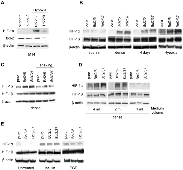
Background: Hypoxia-Inducible Factor 1 (HIF-1) is a transcription factor that is a critical mediator of the cellular response to hypoxia. Enhanced levels of HIF-1alpha, the oxygen-regulated subunit of HIF-1, is often associated with increased tumour angiogenesis, metastasis, therapeutic resistance and poor prognosis. It is in this context that we previously demonstrated that under hypoxia, bcl-2 protein promotes HIF-1/Vascular Endothelial Growth Factor (VEGF)-mediated tumour angiogenesis.

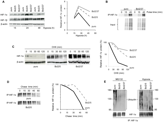
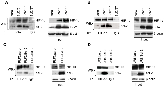
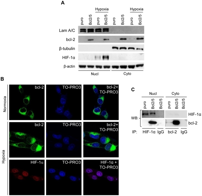
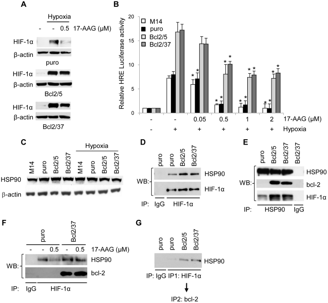
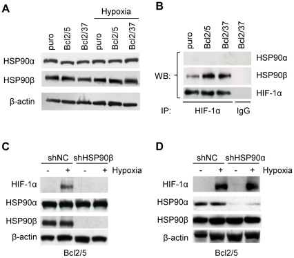
Methodology/principal findings: By using human melanoma cell lines and their stable or transient derivative bcl-2 overexpressing cells, the current study identified HIF-1alpha protein stabilization as a key regulator for the induction of HIF-1 by bcl-2 under hypoxia. We also demonstrated that bcl-2-induced accumulation of HIF-1alpha protein during hypoxia was not due to an increased gene transcription or protein synthesis. In fact, it was related to a modulation of HIF-1alpha protein expression at a post-translational level, indeed its degradation rate was faster in the control lines than in bcl-2 transfectants. The bcl-2-induced HIF-1alpha stabilization in response to low oxygen tension conditions was achieved through the impairment of ubiquitin-dependent HIF-1alpha degradation involving the molecular chaperone HSP90, but it was not dependent on the prolyl hydroxylation of HIF-1alpha protein. We also showed that bcl-2, HIF-1alpha and HSP90 proteins form a tri-complex that may contribute to enhancing the stability of the HIF-1alpha protein in bcl-2 overexpressing clones under hypoxic conditions. Finally, by using genetic and pharmacological approaches we proved that HSP90 is involved in bcl-2-dependent stabilization of HIF-1alpha protein during hypoxia, and in particular the isoform HSP90beta is the main player in this phenomenon.

Conclusions/significance: We identified the stabilization of HIF-1alpha protein as a mechanism through which bcl-2 induces the activation of HIF-1 in hypoxic tumour cells involving the beta isoform of molecular chaperone HSP90.


Conflict of interest statement

Competing Interests: The authors have declared that no competing interests exist.

Figures

Figure 1
Figure 1. bcl-2 modulation regulates HIF-1α protein expression in conditions strictly dependent on oxygen avaibility.
(A) Western blot analysis of HIF-1α and bcl-2 protein expression in total extracts of M14 cells transfected with siRNA targeting bcl-2 mRNA (si-bcl-2) or with a control scrambled si-RNA (si-contr) and then exposed to normoxia or hypoxia for 24 h. (B) Western blot analysis of HIF-1α and HIF-1β protein expression in total extracts of M14 control (puro) and bcl-2 stably overexpressing (Bcl2/5, Bcl2/37) cells plated under low (sparse) or high (dense) cell density conditions, or cultured under normoxia for 4 days or under hypoxia for 24 h. Western blot analysis of HIF-1α and HIF-1β protein expression in total extracts of the cells plated under high cell density conditions and (C) exposed to 24 h shaking or (D) cultured with different volumes of medium. (E) Western blot analysis of HIF-1α and HIF-1β protein expression in total extracts of cells exposed to Insulin (100 nM) or Epidermal Growth Factor (EGF, 20 ng/ml) for 24 h. (A–E) β-actin protein amounts are used to check equal loading and transfer of proteins. Western blot analyses representative of two independent experiments with similar results are shown.
Figure 2
Figure 2. bcl-2 promotes HIF-1α protein stability preventing its ubiquitin-mediated degradation.
(A) Western blot analysis (left panel) and quantification (right panel) of HIF-1α protein expression in M14 control (puro) and bcl-2 stably overexpressing (Bcl2/5, Bcl2/37) clones exposed to hypoxia for the indicated time. (B) Pulse analysis of HIF-1α protein synthesis rate in cells exposed to [35S]–labeled methionine and cysteine for the indicated time. (C) Western blot analysis (left panel) and quantification (right panel) of HIF-1α protein expression in cells exposed to hypoxia for 24 h and then treated with Cyclohexamide (CHX, 50 µg/ml) for the indicated time. (D) Pulse-chase analysis of HIF-1α protein (left panel) and quantification (right panel) in cells plated under dense conditions, pulsed for 45 min with [35S]–labeled methionine and cysteine and chased for the indicated time. (B,D) Whole cell lysates were immunoprecipitated (IP) with anti-HIF-1α antibody and subjected to SDS-PAGE. (E) Western blot analysis of HIF-1α ubiquitination in the cells exposed to MG132 (10 µM, 6 h) or to hypoxia for 24 h. Whole cell lysates were immunoprecipitated (IP) with anti-HIF-1α antibody and then the Western blot analysis was performed using anti-Ubiquitin antibody. (A,C) β-actin protein amounts are used to check equal loading and transfer of proteins and to quantify relative HIF-1α protein levels. (A–E) Western blot, pulse and pulse-chase analyses representative of two independent experiments with similar results are shown. (A,C,D) Densitometric analysis (right panel) of the relative Western blot or Pulse-chase analysis (left panel) was performed using Molecular Analyst Software and normalized with relative controls.
Figure 3
Figure 3. bcl-2 interacts with HIF-1α.
(A) Analysis of HIF-1α/bcl-2 protein interaction in M14 control (puro) and stably bcl-2 overexpressing (Bcl2/5, Bcl2/37) clones exposed to hypoxia for 24 h. Whole cell lysates were immunoprecipitated (IP) with anti-bcl-2 or control (IgG) antibodies and then the Western blot analysis was performed using anti-HIF-1α and anti-bcl-2 antibodies. Analysis of HIF-1α/bcl-2 protein interaction in (B) M14 control (puro) and stably bcl-2 overexpressing (Bcl2/5, Bcl2/37) clones or (C,D) in PLF2 and JR8 control cells (PLF2/puro, JR8/puro) and stably bcl-2 overexpressing (PLF2/Bcl-2, JR8/Bcl-2) cells, exposed to MG132 (10 µM, 6 h). Whole cell lysates were immunoprecipitated with anti-HIF-1α or control (IgG) antibodies and then the Western blot analysis was performed using anti-HIF-1α and anti-bcl-2 antibodies. (A–D) β-actin protein amounts are used to check equal loading and transfer of proteins. Western blot analyses representative of two independent experiments with similar results are shown.
Figure 4
Figure 4. bcl-2 interacts with HIF-1α in the nucleus.
(A) Western blot analysis of bcl-2 and HIF-1α protein expression in nuclear (Nucl) and cytoplasmic (Cyto) protein extracts of M14 control (puro) and bcl-2 stably overexpressing (Bcl2/5) clones exposed to hypoxia or to normoxia for 24 h. LaminA/C (Lam A/C) and β-tubulin were used as markers for nuclear and cytoplasmic fraction, respectively. β-actin protein amounts are used to check equal loading and transfer of proteins. (B) Confocal laser scanning microscopy of immunofluorescence staining performed on Bcl2/5 stably overexpressing clone exposed to hypoxia or to normoxia for 24 h. Fixed cells were labelled with anti-bcl-2 (green) or anti-HIF-1α (red) antibodies. Nuclei were visualized using TO-PRO3® staining (blue). (C) Analysis of HIF-1α/bcl-2 interaction in Bcl2/5 stably overexpressing clone exposed to hypoxia for 24 h. Nuclear (Nucl) and cytoplasmic (Cyto) protein extracts were immunoprecipitated (IP) with anti-HIF-1α or anti-bcl-2, respectively, or control antibody (IgG) and then the Western blot analysis was performed using anti-bcl-2 or anti-HIF-1α antibodies. (A–C) Western blot and confocal analyses representative of two independent experiments with similar results are shown.
Figure 5
Figure 5. HIF-1α prolyl hydroxylation is not required for bcl-2-induced increase of HIF-1α expression and HIF-1 activity in hypoxia.
(A) Western blot analysis of HIF-1α, bcl-2 and PHD2 protein expression and (B) HRE-dependent transcriptional activity in M14 cells stably expressing HA-HIF-1α wild-type (HIF1α wt) or mutated (HIF1α PP/AA), after transiently transfection with control vector (empty), bcl-2 or PHD2 expressing vectors, and then exposure to hypoxia for 24 h. (A) β-actin protein amounts are used to check equal loading and transfer of proteins. Western blot analyses representative of two independent experiments with similar results are shown. (B) Relative luciferase activity of each sample were normalized to the control vector transfected cells. Results represent the mean ± SD of 3 independent experiments performed in triplicate, * p≤0.01.
Figure 6
Figure 6. bcl-2 forms a complex with HSP90 and HIF-1α proteins.
(A) Western blot analysis of HIF-1α protein expression in M14 control cells (puro) and bcl-2 stably overexpressing (Bcl2/5, Bcl2/37) clones treated with 17-AAG under hypoxia or exposed to normoxia for 24 h. (B) HRE-dependent transcriptional activity in the cells treated with 17-AAG from 0.05 to 2 µM under hypoxia or exposed to normoxia for 24 h. Relative luciferase activity of each sample was normalized to untreated cells exposed to normoxic conditions. Results represent the average ± SD of 3 independent experiments performed in triplicate. p values were calculated relative to untreated cells exposed to hypoxic conditions, *p≤0.01. (C) Western blot analysis of HSP90 protein expression in parental M14 cells, control (puro) and bcl-2 stably overexpressing (Bcl2/5, Bcl2/37) clones. (D) Analysis of HIF-1α/HSP90 interaction in the cells exposed to hypoxia for 24 h. Whole cell lysates were immunoprecipitated (IP) with anti-HIF-1α or control (IgG) antibodies and then the Western blot analysis was performed using anti-HSP90 and anti-HIF-1α antibodies. (E) Analysis of HSP90/HIF-1α and HSP90/bcl-2 interactions in the cells exposed to hypoxia for 24 h. Cell lysates were immunoprecipitated (IP) with anti-HSP90 or control (IgG) antibodies and then the Western blot analysis was performed using anti-HIF-1α, anti-bcl-2 and anti-HSP90 antibodies. (F) Analysis of HIF-1α/HSP90 and HIF-1α/bcl-2 interactions in the cells treated with 0.5 µM 17-AAG for 24 h under hypoxia. Whole cell lysates were immunoprecipitated (IP) with anti-HIF-1α antibody and then the Western blot analysis was performed using specific anti-HSP90 and bcl-2 antibodies. (G) Analysis of HSP90/HIF-1α/bcl-2 protein complex in the cells exposed to hypoxia for 24 h. Whole cell lysates were sequentially immunoprecipitated with anti-HIF-1α (IP1) and anti-bcl-2 antibodies (IP2) and then the Western blot analysis was performed using anti-HSP90 antibody. (A,C) β-actin protein amounts are used to check equal loading and transfer of proteins.
Figure 7
Figure 7. HSP90β is the mediator of HIF-1α induction by bcl-2 under hypoxic conditions.
(A) Western blot analysis of HSP90α and HSP90β protein expression in M14 control (puro) and bcl-2 stably overexpressing (Bcl2/5, Bcl2/37) clones exposed to hypoxia or to normoxia for 24 h. (B) Analysis of HSP90α/HIF-1α and HSP90β/HIF-1α interactions in the cells exposed to hypoxia for 24 h. Protein extracts were immunoprecipitated (IP) with anti-HIF-1α and then Western blot analysis was performed using anti-HSP90α and anti-HSP90β antibodies. (C,D) Western blot analysis of HIF-1α, HSP90α and HSP90β protein expression in bcl-2 stably overexpressing cells transiently transfected with short hairpin construct targeting HSP90β (shHSP90β), HSP90α (shHSP90α) or with control vector (shNC) and exposed to hypoxia or to normoxia for 24 h. (A,C,D) β-actin protein amounts are used to check equal loading and transfer of proteins. (A–D) Western blot analyses representative of two independent experiments with similar results are shown.

Similar articles

Cited by

References

    1. Semenza GL. Targeting HIF-1 for cancer therapy. Nat Rev Cancer. 2003;3:721–732. - PubMed
    1. Liu YV, Baek JH, Zhang H, Diez R, Cole RN, et al. RACK1 competes with HSP90 for binding to HIF-1alpha and is required for O(2)-independent and HSP90 inhibitor-induced degradation of HIF-1alpha. Mol Cell. 2007;25:207–217. - PMC - PubMed
    1. Jeong JW, Bae MK, Ahn MY, Kim SH, Sohn TK, et al. Regulation and destabilization of HIF-1alpha by ARD1-mediated acetylation. Cell. 2002;111:709–720. - PubMed
    1. Cho H, Ahn DR, Park H, Yang EG. Modulation of p300 binding by posttranslational modifications of the C-terminal activation domain of hypoxia-inducible factor-1alpha. FEBS Lett. 2007;581:1542–1548. - PubMed
    1. Bilton R, Trottier E, Pouyssegur J, Brahimi-Horn MC. ARDent about acetylation and deacetylation in hypoxia signalling. Trends Cell Biol. 2006;16:616–621. - PubMed

Publication types

MeSH terms