. 2010 Jul 27;4(7):e759.
doi: 10.1371/journal.pntd.0000759.

Development and validation of a quantitative, high-throughput, fluorescent-based bioassay to detect schistosoma viability

Affiliations

Development and validation of a quantitative, high-throughput, fluorescent-based bioassay to detect schistosoma viability

Emily Peak et al. PLoS Negl Trop Dis. .

Abstract

Background: Schistosomiasis, caused by infection with the blood fluke Schistosoma, is responsible for greater than 200,000 human deaths per annum. Objective high-throughput screens for detecting novel anti-schistosomal targets will drive 'genome to drug' lead translational science at an unprecedented rate. Current methods for detecting schistosome viability rely on qualitative microscopic criteria, which require an understanding of parasite morphology, and most importantly, must be subjectively interpreted. These limitations, in the current state of the art, have significantly impeded progress into whole schistosome screening for next generation chemotherapies.

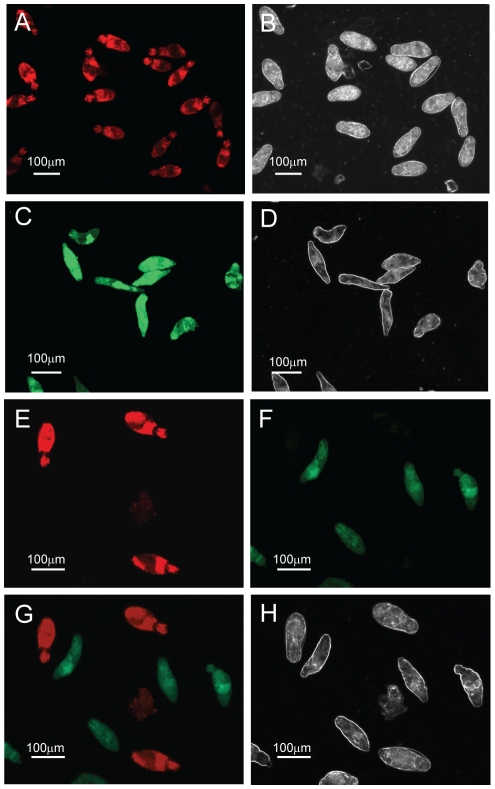
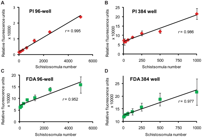
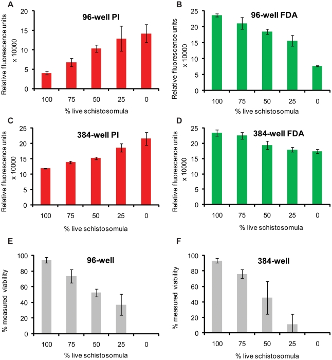
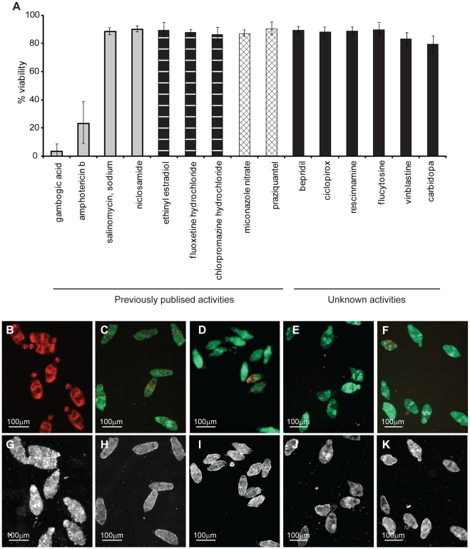
Methodology/principal findings: We present here a microtiter plate-based method for reproducibly detecting schistosomula viability that takes advantage of the differential uptake of fluorophores (propidium iodide and fluorescein diacetate) by living organisms. We validate this high-throughput system in detecting schistosomula viability using auranofin (a known inhibitor of thioredoxin glutathione reductase), praziquantel and a range of small compounds with previously-described (gambogic acid, sodium salinomycin, ethinyl estradiol, fluoxetidine hydrochloride, miconazole nitrate, chlorpromazine hydrochloride, amphotericin b, niclosamide) or suggested (bepridil, ciclopirox, rescinnamine, flucytosine, vinblastine and carbidopa) anti-schistosomal activities. This developed method is sensitive (200 schistosomula/well can be assayed), relevant to industrial (384-well microtiter plate compatibility) and academic (96-well microtiter plate compatibility) settings, translatable to functional genomics screens and drug assays, does not require a priori knowledge of schistosome biology and is quantitative.

Conclusions/significance: The wide-scale application of this fluorescence-based bioassay will greatly accelerate the objective identification of novel therapeutic lead targets/compounds to combat schistosomiasis. Adapting this bioassay for use with other parasitic worm species further offers an opportunity for great strides to be made against additional neglected tropical diseases of biomedical and veterinary importance.


Conflict of interest statement

The authors have declared that no competing interests exist.

Figures

Figure 1
Figure 1. Fluorescein diacetate (FDA) and propidium iodide (PI) can be used to differentially quantify schistosomula viability.
Mechanically-transformed schistosomula were prepared, heat-killed (dead) or left untreated (live) and stained with FDA, PI or both fluorophores according to the Methods . Epi-fluorescent and plane polarized microscopy was used to visualize uptake of fluorophores and examine schistosomula morphology. (A) Dead schistosomula stained with PI and detected by a rhodamine filter (536 nm), (B) Dead schistosomula visualized by polarized light, (C) Live schistosomula stained with FDA and detected by a FITC filter (494 nm), (D) Live schistosomula visualized by polarized light (E) Mixtures of dead and live schistosomula simultaneously stained with PI/FDA and detected by a rhodamine filter, (F) Mixtures of dead and live schistosomula simultaneously stained with PI/FDA and detected by a FITC filter, (G) Differential detection of PI-positive dead and FDA-positive live schistosomula by superimposition of both 536 nm and 494 nm epifluorescent spectra, (H) Differential morphology of dead and live schistosomula detected by polarized light.
Figure 2
Figure 2. A kinetic study of FDA and PI emission reveals the optimal timeframes to collect fluorescent signals from stained schistosomula samples.
Mechanically-transformed schistosomula were cultured for 24 hr, heat killed (dead) or left untreated (live), simultaneously stained with PI and FDA and subjected to fluorescent readings at minute intervals. PI fluorescence (544 nm) was measured over 120 min whereas FDA fluorescence (485 nm) was measured over 51 min. (A) PI fluorescence data collected from a 96-well microtiter plate, (B) PI data collected from a 384-well microtiter plate, (C) FDA data collected from a 96-well microtiter plate and (D) FDA data collected from a 384-well microtiter plate. All fluorescent readings were obtained from a BMG Labtech Polarstar Omega microtiter plate reader. Red lines represent fluorescent data originating from wells containing dead schistosomula, green lines represent fluorescent data originating from wells containing live schistosomula, brown lines represent fluorescent data from wells containing an equal number (mixed) of dead and live schistosomula and dotted lines represent fluorescent data originating from wells containing no schistosomula (media). * Indicates chosen time point for collecting PI data and ♦ indicates chosen time point for collecting FDA data in subsequent experiments. All experiments were performed at least three times. Schistosomula were plated at 1000 parasites/well in the 96-well microtiter plate format and 200 parasites/well in the 384-well microtiter plate format.
Figure 3
Figure 3. A schistosomula titration series reveals the optimal number of parasites to be used for PI and FDA fluorescent detection in both 96-well and 384-well microtiter plates.
Mechanically-transformed schistosomula were cultured for 24 hr, heat killed (dead) or left untreated (live) and stained with either PI (dead parasites) or FDA (live parasites). (A) Dead schistosomula were titrated from 5000 to 36 parasites per well (in triplicate) in a 96-well microtiter plate and PI fluorescence (544 nm) obtained at 20 min, (B) Dead schistosomula were titrated from 1000 to 8 parasites per well (in triplicate) in a 384-well microtiter plate and PI fluorescence (544 nm) obtained at 20 min, (C) Untreated, live schistosomula were titrated from 5000 to 36 parasites per well (in triplicate) in a 96-well microtiter plate and FDA fluorescence (485 nm) measured at 5 min, (D) Untreated, live schistosomula were titrated from 1000 to 8 parasites per well (in triplicate) in a 384-well microtiter plate and FDA fluorescence (485 nm) measured at 5 min. All fluorescent readings were collected from a BMG Labtech Polarstar Omega microtiter plate reader. These results are representative of two independent experiments.
Figure 4
Figure 4. Dual FDA and PI staining of schistosomula samples allows for fluorescent quantification of parasite viability.
Mechanically-transformed schistosomula were cultured for 24 hr, heat killed (dead) or left untreated (live), distributed into wells at pre-defined percentages (100% live, 75% live, 50% live, 25% live 0% live) and simultaneously stained with PI and FDA. (A) Pre-defined percentages of live and dead schistosomula (1000 parasites per well, in triplicate) distributed into a 96-well microtiter plate with PI fluorescence (544 nm) measured at 20 min, (B) Pre-defined percentages of live and dead schistosomula (1000 parasites per well, in triplicate) distributed into a 96-well microtiter plate with FDA fluorescence (485 nm) measured at 5 min, (C) Pre-defined percentages of live and dead schistosomula (200 parasites per well, in triplicate) distributed into a 384-well microtiter plate with PI fluorescence (544 nm) detected at 20 min, (D) Pre-defined percentages of live and dead schistosomula (200 parasites per well, in triplicate) distributed into a 384-well microtiter plate with FDA fluorescence (485 nm) detected at 5 min. (E) Schistosomula viability calculated from fluorescent data displayed in A and B, (F) Schistosomula viability calculated from fluorescent data presented in C and D. Formula for viability calculations is indicated in the Methods section. These results are representative of experiments performed three times.
Figure 5
Figure 5. Dual-fluorescence viability determination of auranofin treated schistosomula.
Mechanically-transformed schistosomula were cultured for 24 hr, incubated with different concentrations of auranofin for an additional 24 hr, washed and subsequently co-stained with both PI and FDA. PI- (544 nm, collected at 20 min) and FDA- (485 nm, collected at 5 min) fluorescence intensity units were converted into viability measures according to the formula described in the Methods . (A) Dose-dependent anti-schistosomula effect of auranofin as indicated by % viability. Treatments showing statistically significant differences in viability when compared to untreated (live) schistosomula are indicated with * (p<0.05) or ** (p<0.001). (B) Auranofin dose-response curve by which an LD50 was calculated by plotting the probit transformation of the % viability to the Log10 transformation of auranofin concentration. Dotted line indicates the average LD50 value calculated from three replicates. (C) Light microscope image of schistosomula treated with 10µM auranofin for 24 hr. (D) Light microscope image of untreated schistosomula. (E) Epi-fluorescent and (F) plane polarized light micrograph of schistosomula treated with 10µM auranofin for 24 hr, then incubated with both PI and FDA. (G) Epi-fluorescent and (H) plane polarized light micrograph of schistosomula treated with 1µM auranofin for 24 hr, then incubated with PI and FDA. ‘Dead’ represents schistosomula killed by heat-treatment and ‘0 µM’ auranofin represents schistosomula incubated with 1% (v/v) DMSO (auranofin solvent). These results are representative of experiments performed three times.
Figure 6
Figure 6. Application of the dual-fluorescent viability assay for determining the anti-schistosomula effect of selected compounds with previously-described or unknown activities.
Mechanically-transformed schistosomula were cultured for 24 hr, incubated with compounds (10 µM) for an additional 24 hr, washed and subsequently co-stained with both PI and FDA. PI- (544 nm, collected at 20 min) and FDA- (485 nm, collected at 5 min) fluorescence intensity units were converted into viability measures according to the formula described in the Methods . (A) Calculated schistosomula viability in response to each compound tested (further details can be found in Dataset S1). Representative epi-fluorescent and plane polarized microscope images of schistosomula treated with (B and G) gambogic acid, (C and H) sodium salinomycin, (D and I) niclosamide, (E and J) praziquantel and (F and K) ciclopirox are indicated. Compounds designated as having ‘previously published activities’ (grey histograms – death, vertical lines within histograms – overactive, hatched lines within histograms – rounded) were selected from those described by Abdulla et al. while those compounds indicated as having ‘unknown activities’ (black histograms) were selected from Berriman et al. . These results are representative of two independent experiments.

References

    1. King CH, Dickman K, Tisch DJ. Reassessment of the cost of chronic helmintic infection: a meta-analysis of disability-related outcomes in endemic schistosomiasis. J Lancet. 2005;365:1561–1569. - PubMed
    1. Fenwick A, Savioli L, Engels D, Robert Bergquist N, Todd MH. Drugs for the control of parasitic diseases: current status and development in schistosomiasis. Trends Parasitol. 2003;19:509–515. - PubMed
    1. Berriman M, Haas BJ, LoVerde PT, Wilson RA, Dillon GP, et al. The genome of the blood fluke Schistosoma mansoni. Nature. 2009;460:352–358. - PMC - PubMed
    1. Brindley PJ, Pearce EJ. Genetic manipulation of schistosomes. Int J for Parasitol. 2007;37:465–473. - PubMed
    1. Hokke CH, Fitzpatrick JM, Hoffmann KF. Integrating transcriptome, proteome and glycome analyses of Schistosoma biology. Trends Parasitol. 2007;23:165–174. - PubMed

Publication types

MeSH terms