. 2010 Jul 23;5(7):e11765.
doi: 10.1371/journal.pone.0011765.

Cholesterol crystals activate the NLRP3 inflammasome in human macrophages: a novel link between cholesterol metabolism and inflammation

Affiliations

Cholesterol crystals activate the NLRP3 inflammasome in human macrophages: a novel link between cholesterol metabolism and inflammation

Kristiina Rajamäki et al. PLoS One. .

Abstract

Background: Chronic inflammation of the arterial wall is a key element in the pathogenesis of atherosclerosis, yet the factors that trigger and sustain the inflammation remain elusive. Inflammasomes are cytoplasmic caspase-1-activating protein complexes that promote maturation and secretion of the proinflammatory cytokines interleukin(IL)-1beta and IL-18. The most intensively studied inflammasome, NLRP3 inflammasome, is activated by diverse substances, including crystalline and particulate materials. As cholesterol crystals are abundant in atherosclerotic lesions, and IL-1beta has been linked to atherogenesis, we explored the possibility that cholesterol crystals promote inflammation by activating the inflammasome pathway.

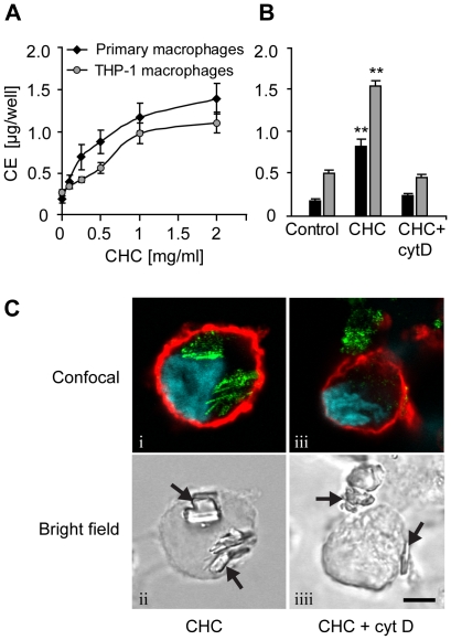
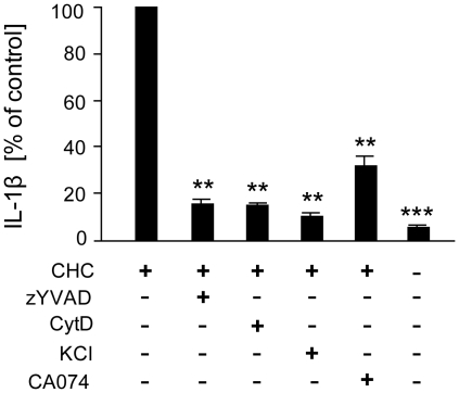
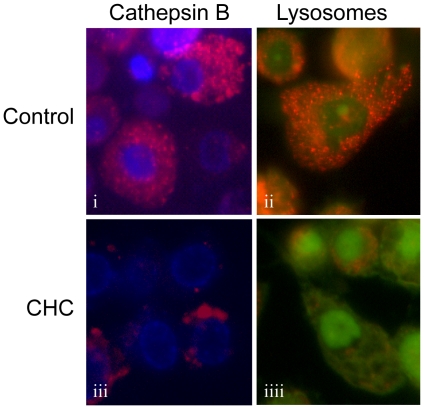
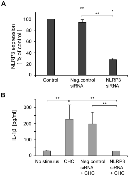
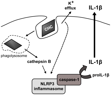
Principal findings: Here we show that human macrophages avidly phagocytose cholesterol crystals and store the ingested cholesterol as cholesteryl esters. Importantly, cholesterol crystals induced dose-dependent secretion of mature IL-1beta from human monocytes and macrophages. The cholesterol crystal-induced secretion of IL-1beta was caspase-1-dependent, suggesting the involvement of an inflammasome-mediated pathway. Silencing of the NLRP3 receptor, the crucial component in NLRP3 inflammasome, completely abolished crystal-induced IL-1beta secretion, thus identifying NLRP3 inflammasome as the cholesterol crystal-responsive element in macrophages. The crystals were shown to induce leakage of the lysosomal protease cathepsin B into the cytoplasm and inhibition of this enzyme reduced cholesterol crystal-induced IL-1beta secretion, suggesting that NLRP3 inflammasome activation occurred via lysosomal destabilization.

Conclusions: The cholesterol crystal-induced inflammasome activation in macrophages may represent an important link between cholesterol metabolism and inflammation in atherosclerotic lesions.


Conflict of interest statement

Competing Interests: The authors have declared that no competing interests exist.

Figures

Figure 1
Figure 1. Macrophages accumulate cholesteryl esters when incubated with cholesterol crystals (CHCs).
(A) Primary macrophages and THP-1 macrophages were incubated with 0.1–2 mg/ml CHCs and cholesteryl esters (CE) were measured from cellular lipid extracts by TLC. (B) Cytochalasin D (cytD; 2 µM) was employed to block cytoskeletal movements during a 16 h incubation of primary and THP-1 macrophages with CHCs (0.5 and 1.0 mg/ml, respectively). (C) THP-1 macrophages treated with CHCs ± cytD were stained with fluorophore-conjugated cholera toxin subunit B (cell membrane; red) and Hoechst (nuclei; blue). The cells were imaged using confocal fluorescence microscopy, combined with detection of CHCs by confocal reflection of the 488 nm laser line (green) (panels i, iii). CHCs are indicated by arrows in the bright field panels (ii, iiii). Scale bar 5 µm. Each experiment was performed ≥4 times. The data are means ± s.e.m. ** = p<0.01, compared to the control cells.
Figure 2
Figure 2. Monocytes and macrophages respond to cholesterol crystals (CHCs) by IL-1β secretion.
Primary monocytes (A), primary macrophages (B,D,E), and THP-1 macrophages (C) were incubated with CHCs for 16 h, 24 h, and 8 h, respectively. LPS (1 µg/ml) was used as a co-stimulant for the primary cells. Concentrations of cytokines were subsequently determined from cell culture supernatants. The inset in 2C verifies the presence of mature 17 kDa IL-1β in cell culture supernatants of THP-1 macrophages by Western blotting. The data are means ± s.e.m. from ≥4 experiments. * = p<0.05 and ** = p<0.01, compared to untreated cells.
Figure 3
Figure 3. Analysis of inflammasome-related gene expression after exposure of cells to cholesterol crystals (CHCs).
Primary macrophages from 3 donors were incubated for 1–9 h with (A) 0.5 mg/ml CHCs and 1 µg/ml LPS, (B) with LPS alone, or (C) with CHCs alone. Untreated control cells were included at each time point. After the incubation, mRNA levels were determined by real-time quantitative RT-PCR. The data are expressed as mean fold changes ± s.e.m. relative to the untreated cells at each time point.
Figure 4
Figure 4. Mechanism of cholesterol crystal (CHC)-induced IL-1β secretion.
THP-1 macrophages were incubated with CHCs in the absence or presence of caspase-1 inhibitor zYVAD-fmk (25 µM), cytochalasin D (2 µM), KCl (130 mM), or cathepsin B inhibitor CA-074Me (10 µM). After the incubation, cell culture supernatants were analyzed for IL-1β (average response to CHCs 662 pg/ml). The data are means ± s.e.m. from ≥5 experiments. ** = p<0.01 and *** = p<0.001, compared with CHC-treated cells.
Figure 5
Figure 5. Cholesterol crystals (CHCs) cause destabilization of lysosomes and leakage of cathepsin B into the cytoplasm.
CHC-treated or untreated live THP-1 macrophages were stained with cathepsin B substrate z-Arg-Arg-cresyl violet (panels i, iii) or with acridine orange (panels ii, iiii). The fluorescent cresyl violet group of z-Arg-Arg-cresyl violet is dequenched upon cleavage of one or both of the arginines by cathepsin B. Acridine orange aggregates in the acidic pH of lysosomes, which changes the fluorescence emission of the dye from green to red. The images are representative of 3 experiments.
Figure 6
Figure 6. Silencing of NLRP3 attenuates cholesterol crystal (CHC)-induced IL-1β secretion.
(A) NLRP3 mRNA levels were reduced by 72% after treatment of THP-1 macrophages with NLRP3-targeted small interfering RNA (siRNA). (B) CHC-induced IL-1β secretion was abolished after treatment of cells with NLRP3 siRNA, whereas treatment of cells with negative control siRNA had no effect. The data are means ± s.e.m. from 5 (A) and 3 (B) experiments. ** = p<0.01.
Figure 7
Figure 7. Proposed mechanism of cholesterol crystal (CHC)-induced inflammasome activation.
CHCs are phagocytosed by macrophages, causing lysosomal destabilization and leakage of cathepsin B to cytoplasm, where the enzyme indirectly activates the NLRP3 inflammasome. Lowering of intracellular potassium concentration, stemming from potassium efflux caused by phagocytosed CHCs, is also required for NLRP3 activation.

References

    1. Galkina E, Ley K. Immune and inflammatory mechanisms of atherosclerosis. Annu Rev Immunol. 2009;27:165–197. - PMC - PubMed
    1. Tedgui A, Mallat Z. Cytokines in atherosclerosis: Pathogenic and regulatory pathways. Physiol Rev. 2006;86(2):515–581. - PubMed
    1. Galea J, Armstrong J, Gadsdon P, Holden H, Francis SE, et al. Interleukin-1 beta in coronary arteries of patients with ischemic heart disease. Arterioscler Thromb Vasc Biol. 1996;16(8):1000–1006. - PubMed
    1. Kirii H, Niwa T, Yamada Y, Wada H, Saito K, et al. Lack of interleukin-1beta decreases the severity of atherosclerosis in ApoE-deficient mice. Arterioscler Thromb Vasc Biol. 2003;23(4):656–660. - PubMed
    1. Wang X, Feuerstein GZ, Gu JL, Lysko PG, Yue TL. Interleukin-1 beta induces expression of adhesion molecules in human vascular smooth muscle cells and enhances adhesion of leukocytes to smooth muscle cells. Atherosclerosis. 1995;115(1):89–98. - PubMed

Publication types

MeSH terms