Review
. 2010 Dec;33(6):1079-87.
doi: 10.1007/s00270-010-9943-6. Epub 2010 Jul 29.

Literature review of the role of ultrasound, computed tomography, and transcatheter arterial embolization for the treatment of traumatic splenic injuries

Affiliations
Review

Literature review of the role of ultrasound, computed tomography, and transcatheter arterial embolization for the treatment of traumatic splenic injuries

Cornelis H van der Vlies et al. Cardiovasc Intervent Radiol. 2010 Dec.

Abstract

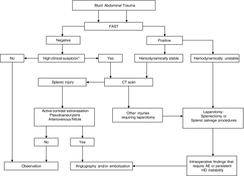
Introduction: The spleen is the second most frequently injured organ following blunt abdominal trauma. Trends in management have changed over the years. Traditionally, laparotomy and splenectomy was the standard management. Presently, nonoperative management (NOM) of splenic injury is the most common management strategy in hemodynamically stable patients. Splenic injuries can be managed via simple observation (OBS) or with angiography and embolization (AE). Angio-embolization has shown to be a valuable alternative to observational management and has increased the success rate of nonoperative management in many series.

Diagnostics: Improved imaging techniques and advances in interventional radiology have led to a better selection of patients who are amenable to nonoperative management. Despite this, there is still a lot of debate about which patients are prone to NOM.

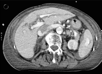
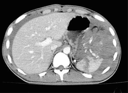
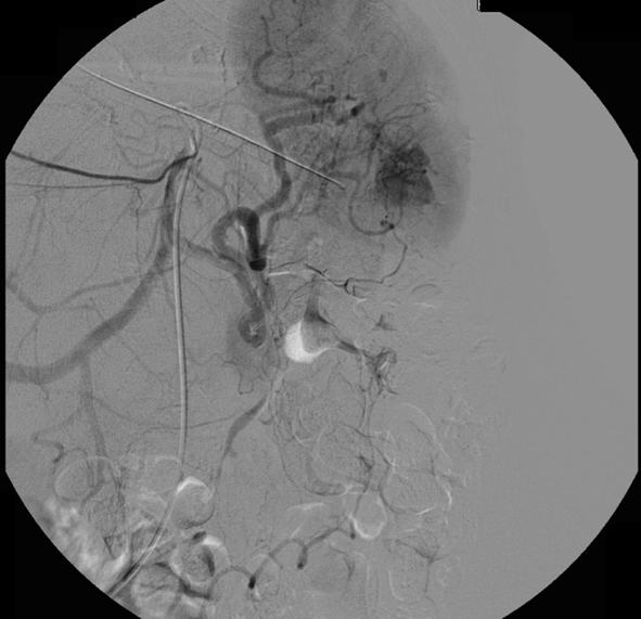
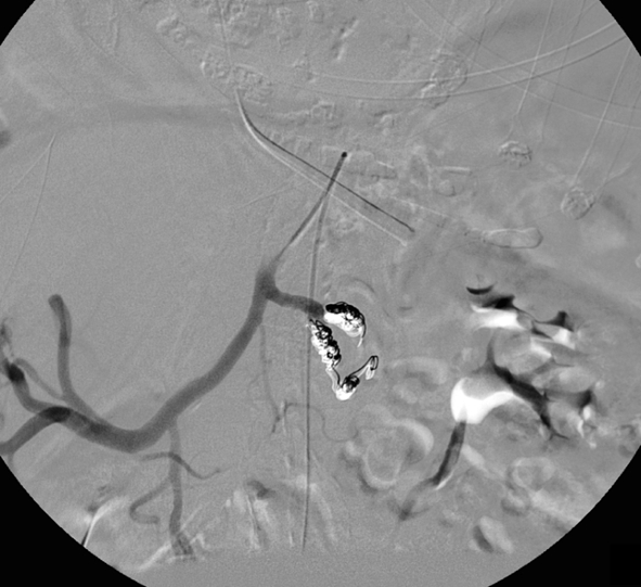
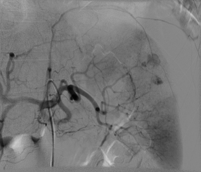
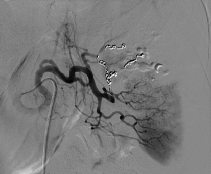
Angiography and embolization: The optimal patient selection is still a matter of debate and the role of CT and angio-embolization has not yet fully evolved. We discuss the role of sonography and CT features, such as contrast extravasation, pseudoaneurysms, arteriovenous fistulas, or hemoperitoneum, to determine the optimal patient selection for angiography and embolization. We also review the efficiency, technical considerations (proximal or selective embolization), logistics, and complication rates of AE for blunt traumatic splenic injuries.


Figures

Fig. 1
Fig. 1
Hemodynamically stable patient (patient A) with blunt abdominal trauma after fall from horse. CT with intravenous contrast shows small amount of hemoperitoneum around the spleen and a contrast “blush,” which is confined to the splenic parenchyma (AAST grade 3, Baltimore grade 4a)
Fig. 2
Fig. 2
Hemodynamically stable patient (patient B) with blunt abdominal trauma after motor vehicle accident. CT with intravenous contrast shows hemoperitoneum, fractured spleen with large hematoma and extravasation of contrast medium into the abdominal cavity (AAST grade 4, Baltimore grade 4b)
Fig. 3
Fig. 3
Flowchart for the diagnostic and therapeutic management of blunt splenic injury. *Abnormalities in physical examination of the abdomen, pelvis, or lumbar spine, base excess <−3, systolic blood pressure <90 mmHg, long bone fractures [27]
Fig. 4
Fig. 4
Same patient (patient A) as in Fig. 1. Selective digital subtraction angiogram of the celiac axis showing the intraparenchymal contrast “blush” in the spleen. Note tortuous anatomy of the splenic artery
Fig. 5
Fig. 5
Same patient (patient A) as Figs. 1 and 4. Selective embolization was not possible as a result of the tortuous anatomy and proximal coil embolization of the splenic artery was performed. Check angiogram after embolization shows occlusion of the proximal splenic artery. Perfusion of the spleen by collaterals is not shown in this image
Fig. 6
Fig. 6
Same patient (patient B) as in Fig. 2. Selective digital subtraction angiogram of the celiac axis showing multiple areas of contrast extravasation from peripheral branches of the splenic artery
Fig. 7
Fig. 7
Same patient (patient B) as in Figs. 2 and 6. Check angiogram after selective coil embolization of an interpolar branch of the splenic artery. Contrast extravasation is no longer seen, and there is good perfusion of the remainder of the spleen

References

    1. Smith J, Caldwell E, D’Amours S, Jalaludin B, Sugrue M. Abdominal trauma: a disease in evolution. ANZ J Surg. 2005;75:790–794. doi: 10.1111/j.1445-2197.2005.03524.x. - DOI - PubMed
    1. Malangoni MA, Cue JI, Fallat ME, Willing SJ, Richardson JD. Evaluation of splenic injury by computed tomography and its impact on treatment. Ann Surg. 1990;211:592–597. doi: 10.1097/00000658-199005000-00009. - DOI - PMC - PubMed
    1. Gopal V, Bisno AL. Fulminant pneumococcal infections in “normal” asplenic hosts. Arch Int Med. 1977;137:1526–1530. doi: 10.1001/archinte.137.11.1526. - DOI - PubMed
    1. Holdsworth RJ, Irving AD, Cuschieri A. Postsplenectomy sepsis and its mortality rate: actual versus perceived risks. Br J Surg. 1991;78:1031–1038. doi: 10.1002/bjs.1800780904. - DOI - PubMed
    1. Pachter HL, Hofstetter SR, Spencer FC. Evolving concepts in splenic surgery: splenorrhaphy versus splenectomy and postsplenectomy drainage: experience in 105 patients. Ann Surg. 1981;194:262–269. doi: 10.1097/00000658-198109000-00003. - DOI - PMC - PubMed